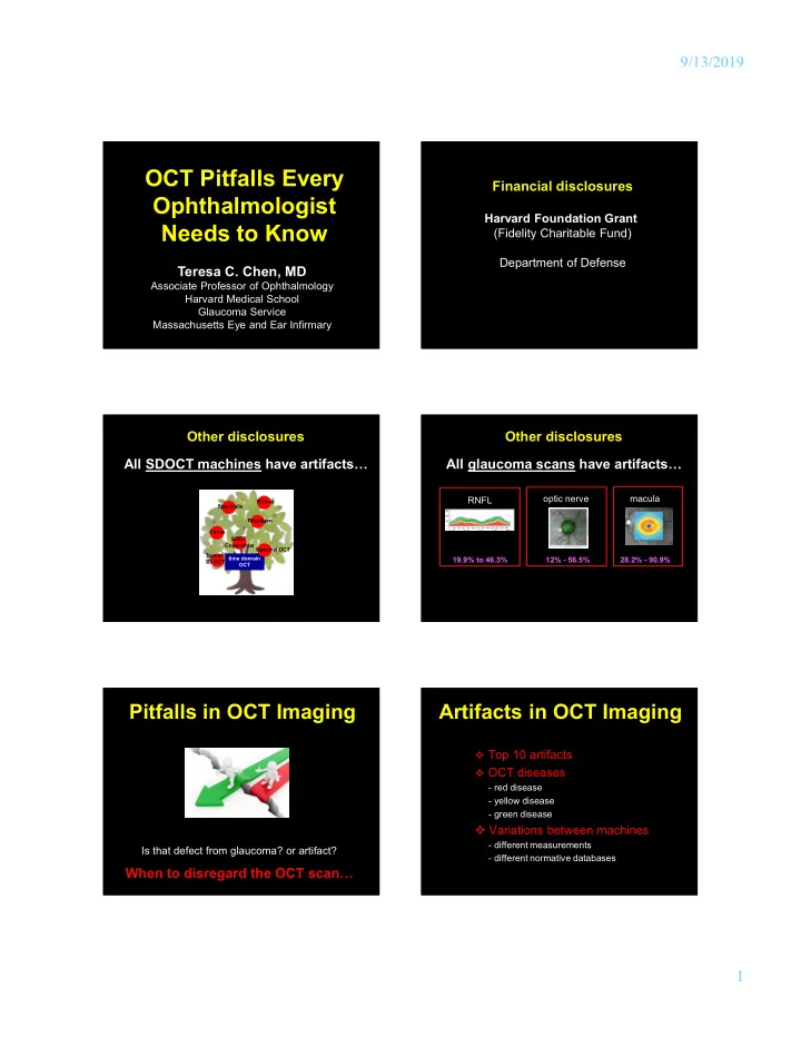

9/13/2019 OCT Pitfalls Every Financial disclosures Ophthalmologist Harvard Foundation Grant Needs to Know (Fidelity Charitable Fund) Department of Defense Teresa C. Chen, MD Associate Professor of Ophthalmology Harvard Medical School Glaucoma Service Massachusetts Eye and Ear Infirmary Other disclosures Other disclosures All SDOCT machines have artifacts… All glaucoma scans have artifacts… optic nerve macula RNFL RTVue Spectralis Bioptigen Cirrus SOCT Copernicus Spectral OCT Topcon time domain 19.9% to 46.3% 12% - 56.5% 28.2% - 90.9% 3D OCT OCT Pitfalls in OCT Imaging Artifacts in OCT Imaging Top 10 artifacts OCT diseases - red disease - yellow disease - green disease Variations between machines - different measurements Is that defect from glaucoma? or artifact? - different normative databases When to disregard the OCT scan… 1
9/13/2019 Artifacts in OCT Imaging Artifacts in OCT Imaging Cut edge 83.8 – 87.3% of RNFL and macular (0.17% of RNFL scans)… artifacts were obvious on the printout 10 1,118 patients with Spectralis RNFL Yingna Liu, Huseyin Simavli, Christian Que, Jennifer Rizzo, Edem Tsikata, Rie Maurer, Teresa Chen. Am J Ophthalmol 2015. 213 patients with Spectralis RNFL Steven Mansberger, Shivali Menda, Brad Fortune, Stuart Gardiner, Shaban Demirel. Am J Ophthalmol 2017. Patient Characteristics Associated with Artifacts in Spectralis OCT 277 patients with Spectralis RNFL Imaging of the Retinal Nerve Fiber Layer. Yingna Liu, Huseyin Simavli, Sanjay Asrani, Luma Essaid, Brian Alder, Christian Que, Jennifer Rizzo, Edem Tsikata, Rie Maurer, Teresa Chen. Cecilia Santiago-Turla. JAMA Ophthalmology 2014. Am J Ophthalmol 2015; 159:565-576. Artifacts in OCT Imaging Artifacts in OCT Imaging Motion artifact Incomplete segmentation 9 8 (0.2% of RNFL scans)… (0.6% of RNFL scans)…… Patient Characteristics Associated with Artifacts in Spectralis OCT Patient Characteristics Associated with Artifacts in Spectralis OCT Imaging of the Retinal Nerve Fiber Layer. Yingna Liu, Huseyin Simavli, Imaging of the Retinal Nerve Fiber Layer. Yingna Liu, Huseyin Simavli, Christian Que, Jennifer Rizzo, Edem Tsikata, Rie Maurer, Teresa Chen. Christian Que, Jennifer Rizzo, Edem Tsikata, Rie Maurer, Teresa Chen. Am J Ophthalmol 2015; 159:565-576. Am J Ophthalmol 2015; 159:565-576. Artifacts in OCT Imaging Artifacts in OCT Imaging Missing parts PPA-associated error (1.5% of RNFL scans)… (1.2% of RNFL scans)… 7 6 optimal scan circle size Patient Characteristics Associated with Artifacts in Spectralis OCT Patient Characteristics Associated with Artifacts in Spectralis OCT Imaging of the Retinal Nerve Fiber Layer. Yingna Liu, Huseyin Simavli, Imaging of the Retinal Nerve Fiber Layer. Yingna Liu, Huseyin Simavli, Christian Que, Jennifer Rizzo, Edem Tsikata, Rie Maurer, Teresa Chen. Christian Que, Jennifer Rizzo, Edem Tsikata, Rie Maurer, Teresa Chen. Am J Ophthalmol 2015; 159:565-576. Am J Ophthalmol 2015; 159:565-576. 2
9/13/2019 Artifacts in OCT Imaging Artifacts in OCT Imaging Missing parts Anterior mis-identification of RNFL (1.5% of RNFL scans)… 6 5 (3.2% of RNFL scans)… Patient Characteristics Associated with Artifacts in Spectralis OCT Imaging of the Retinal Nerve Fiber Layer. Yingna Liu, Huseyin Simavli, Eye Wiki by Anjum Cheema, MD and Christian Que, Jennifer Rizzo, Edem Tsikata, Rie Maurer, Teresa Chen. Daniel Moore MD Am J Ophthalmol 2015; 159:565-576. Artifacts in OCT Imaging Artifacts in OCT Imaging Poor signal (5.1% of RNFL scans)… Poor signal (5.1% of RNFL scans)… 4 4 ….due to dry eyes Effect of Corneal Drying on Optical Coherence Patient Characteristics Associated with Artifacts in Spectralis OCT Tomography. Daniel Stein, Gadi Wollstein, Hiroshi Imaging of the Retinal Nerve Fiber Layer. Yingna Liu, Huseyin Simavli, Ishikawa, Ellen Hertzmark, Robert Noecker, Joel Christian Que, Jennifer Rizzo, Edem Tsikata, Rie Maurer, Teresa Chen. Schuman. Ophthalmology 2006; 113: 985-991. Am J Ophthalmol 2015; 159:565-576. Artifacts in OCT Imaging Artifacts in OCT Imaging image quality indices different for different machines Poor signal (5.1% of RNFL scans)… 4 image quality affects RNFL thickness measurements SDOCT Machine Scan Quality Index …..due to lens opacity Cirrus HD-OCT Signal Strength > 6 (max. 10) RTVue Signal Strength Index (SSI) ≥ 30 (max. 100) 3D-OCT Image quality > 45 (max. 160) Spectralis SD-OCT Quality (Q) > 15 Patient Characteristics Associated with Artifacts in Spectralis OCT (max. 40) Imaging of the Retinal Nerve Fiber Layer. Yingna Liu, Huseyin Simavli, Christian Que, Jennifer Rizzo, Edem Tsikata, Rie Maurer, Teresa Chen. Am J Ophthalmol 2015; 159:565-576. 3
9/13/2019 Artifacts in OCT Imaging Artifacts in OCT Imaging Posterior RNFL misidentification Posterior RNFL misidentification 3 3 (7.7% of RNFL scans)…… (7.7% of RNFL scans)…… Beware of the “floor effect”… Spectralis 49.2 microns Cirrus 57 microns RTVue 64.7 microns Patient Characteristics Associated with Artifacts in Spectralis OCT Comprehensive Three-Dimensional Analysis of the Neuroretinal Rim Imaging of the Retinal Nerve Fiber Layer. Yingna Liu, Huseyin Simavli, in Glaucoma.... Edem Tsikata, Ramon Lee, Eric Shieh, Huseyin Christian Que, Jennifer Rizzo, Edem Tsikata, Rie Maurer, Teresa Chen. Simavli, Christian Que, Rong Guo, Ziad Khouier, Johannes de Boer, Am J Ophthalmol 2015; 159:565-576. Teresa Chen. Invest Ophthalmol Vis Sci 2016. Artifacts in OCT Imaging Artifacts in OCT Imaging PVD-associated error PVD-associated error (14.4% of RNFL scans)… 2 2 (14.4% of RNFL scans)… RNFL too “thin”…. RNFL too “thick”…. …or just “right”…. …or just “right”…. Patient Characteristics Associated with Artifacts in Spectralis OCT Patient Characteristics Associated with Artifacts in Spectralis OCT Imaging of the Retinal Nerve Fiber Layer. Yingna Liu, Huseyin Simavli, Imaging of the Retinal Nerve Fiber Layer. Yingna Liu, Huseyin Simavli, Christian Que, Jennifer Rizzo, Edem Tsikata, Rie Maurer, Teresa Chen. Christian Que, Jennifer Rizzo, Edem Tsikata, Rie Maurer, Teresa Chen. Am J Ophthalmol 2015; 159:565-576. Am J Ophthalmol 2015; 159:565-576. Artifacts in OCT Imaging Artifacts in OCT Imaging De-centration 1 1 (27.8% of RNFL scans)…… Effect of Improper Scan Alignment on RNFL Thickness Patient Characteristics Associated with Artifacts in Spectralis OCT Measurements Using Stratus OCT. Gianmarco Vizzeri, Imaging of the Retinal Nerve Fiber Layer. Yingna Liu, Huseyin Simavli, Christopher Bowd, Felipe Medeiros, Robert Weinreb, Linda Christian Que, Jennifer Rizzo, Edem Tsikata, Rie Maurer, Teresa Chen. Zangwill. Journal of Glaucoma 2008; 17: 341-349. Am J Ophthalmol 2015; 159:565-576. 4
9/13/2019 Artifacts in OCT Imaging Artifacts in OCT Imaging De-centration De-centration (27.8% of RNFL scans)…… 1 (27.8% of RNFL scans)…… 1 well-centered well-centered superiorly inferiorly de-centered de-centered Effect of Improper Scan Alignment on RNFL Thickness Effect of Improper Scan Alignment on RNFL Thickness Measurements Using Stratus OCT. Gianmarco Vizzeri, Measurements Using Stratus OCT. Gianmarco Vizzeri, Christopher Bowd, Felipe Medeiros, Robert Weinreb, Linda Christopher Bowd, Felipe Medeiros, Robert Weinreb, Linda Zangwill. Journal of Glaucoma 2008; 17: 341-349. Zangwill. Journal of Glaucoma 2008; 17: 341-349. Artifacts in OCT Imaging Artifacts in OCT Imaging De-centration De-centration 1 1 (27.8% of RNFL scans)…… (27.8% of RNFL scans)…… well-centered well-centered nasally temporally de-centered de-centered Effect of Improper Scan Alignment on RNFL Thickness Measurements Effect of Improper Scan Alignment on RNFL Thickness Measurements Using Stratus OCT. Gianmarco Vizzeri, Christopher Bowd, Felipe Medeiros, Using Stratus OCT. Gianmarco Vizzeri, Christopher Bowd, Felipe Medeiros, Robert Weinreb, Linda Zangwill. Journal of Glaucoma 2008; 17: 341-349. Robert Weinreb, Linda Zangwill. Journal of Glaucoma 2008; 17: 341-349. Artifacts in OCT Imaging Artifacts in OCT Imaging Top Ten Reasons for OCT Artifacts Top 10 artifacts 1.de-centration OCT diseases 2.PVD-associated error 3.posterior RNFL mis-identification - red disease 4.poor signal - yellow disease 5.anterior RNFL mis-identification - green disease 6.missing parts Variations between machines 7.PPA-associated error - different measurements 8.incomplete segmentation - different normative databases 9.motion artifact 10.cut edge 5
9/13/2019 OCT Diseases OCT Diseases red = glaucoma red disease = false positive yellow = maybe glaucoma green = normal Gabriel Chong, Richard Lee. Glaucoma versus red disease: imaging and glaucoma diagnosis. Current Opinion 2012. Etiologies of Red Disease Red Disease from De-centration glaucoma “cured!” vs. “de-centered” TOP 10 ARTIFACTS de-centration PVD-associated error posterior RNFL mis-identification poor signal anterior RNFL mis-identification missing parts PPA-associated error incomplete segmentation motion artifact cut edge Etiologies of Red Disease Red Disease from Myopia -7.0 myope afflicted by NON-GLAUCOMATOUS CAUSES OF RNFL red disease… THINNING: myopia ischemic optic neuropathy panretinal photocoagulation superior segmental optic nerve hyopoplasia (i.e. topless disc appearance) optic nerve hypoplasia arterial and vein occlusions multiple sclerosis and past optic neuritis machine says patient has glaucoma when he does not 6
Recommend
More recommend