OCT: Applications in Laser Vision Correction and Lens-Based - - PowerPoint PPT Presentation

oct applications in laser vision correction and lens
SMART_READER_LITE
LIVE PREVIEW

OCT: Applications in Laser Vision Correction and Lens-Based - - PowerPoint PPT Presentation

12/3/2016 OCT: Applications in Laser Vision Correction and Lens-Based Refractive Surgery I have no disclosures relating to this talk. Julie Schallhorn, MD MS I have received lecture honoraria from Avellino Labs. University of California, San


slide-1
SLIDE 1

12/3/2016 1

OCT: Applications in Laser Vision Correction and Lens-Based Refractive Surgery

Julie Schallhorn, MD MS University of California, San Francisco F.I. Proctor Foundation

I have received lecture honoraria from Avellino Labs. I have no disclosures relating to this talk.

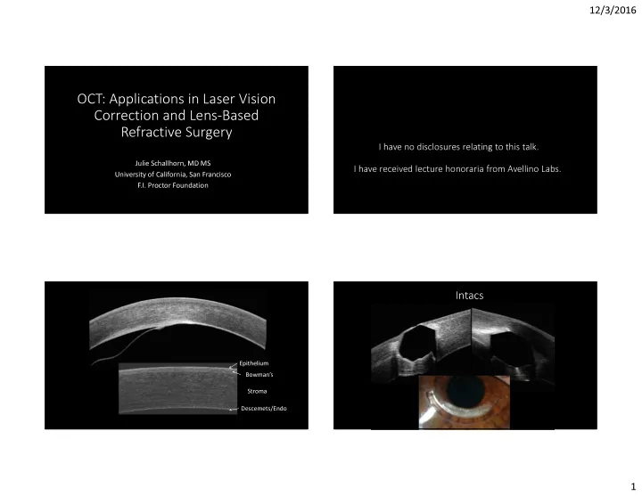
Epithelium Bowman’s Stroma Descemets/Endo

Intacs

slide-2
SLIDE 2

12/3/2016 2

Residual Stromal Bed Measurement Modern Anterior Segment OCT

  • Axial resolution 5-10μm – able to resolve the five layers of the

cornea

  • Images each individual interface in cornea – not just anterior
  • r posterior surface
  • Information about what is going on inside of cornea
  • Subtle perturbations in epithelium, stroma
  • Good news – you probably already have it or can easily get it!

Anterior Segment OCT Options

Device Axial Resolution Field of View Imaging Optovue RTVue/Avanti (CAM Package) 5μm 6mm (9mm in trials) Pachymetry map Epithelial map Line Scans Tomey Casia 2* 10μm 13mm Pachymetry map Topography Line Scans Zeiss Visante 18μm 10mm Pachymetry map Line Scans Zeiss Cirrus (AS Adaptor) 5μm 10mm Pachymetry map Epithelial map* Line Scans Heidelberg Spectralis (AS Adaptor) 4-7μm 6mm Line Scans

* Not FDA approved

Pachymetry Map

Cirrus RTVue

6mm 10mm

slide-3
SLIDE 3

12/3/2016 3

Keratoconic corneas thinner, more focally abnormal Thinning displaced inferior Greater difference between mean thickness and thinnest point

Keratoconus Pachymetry Map

RTVue

Why do I need OCT when I’ve got the Pentacam?

OCT Epithelial Mapping

  • Commercially available on RTVue

with Pachymetry + Cpwr scan software package

  • Ability to differentiate between

stromal and epithelial processes

  • 8 radial scans with automated

boundary detection: tear film to Bowman’s

slide-4
SLIDE 4

12/3/2016 4

Epithelium Map

Epithelial Map

  • Normal Thickness: Central

>Inferior >Superior

  • Epithelium is very sensitive to

underlying corneal curvature- thickness

  • Highly reflective of local

contour changes

OCT & Screening for Ectasia

  • Epithelium thins over steepest portion of cornea and

thickens elsewhere

  • Makes cone appear less steep
  • May be earliest change in ectasia

Keratoconus

Pachymetry Map Epithelium Map Topography

Epithelial Thinning over the Cone Epithelial Thickening around the Cone

Forme Fruste Keratoconus?

slide-5
SLIDE 5

12/3/2016 5

Posterior Float

Forme Fruste Keratoconus?

  • 76 normal eyes/35 KCN eyes
  • Pattern Standard Deviation (PSD) of the Epithelium
  • Indicator of difference from normal epi map
  • PSD ≥ 0.57 – 100% sensitivity and specificity for non-

normal

  • Abnormal in CL warpage, severe dry eye and KCN

Pattern Standard Deviation= 0.57

Forme Fruste Keratoconus?

slide-6
SLIDE 6

12/3/2016 6 Fellow Eye

The epithelium is a very sensitive and specific indicator of early ectasia.

Forme Fruste Keratoconus?

Epithelium Map Topography

Epithelial thickening in area of steepening

Contact Lens Warpage

OCT in Lens-Based Surgery

  • Evaluate the macula
  • Evaluate the macula
  • Evaluate the macula
  • Post-Refractive IOL Calculations
slide-7
SLIDE 7

12/3/2016 7

OCT & IOLs

iolcalc.ascrs.org

  • OCT-based formula had smallest

variance in IOL power and smallest mean error in prediction (0.39D)

  • OCT with similar mean prediction

error to ORA and Hagis-L formulas

On the Horizon

slide-8
SLIDE 8

12/3/2016 8 Epithelial Remodeling, Regression and Refractive Predictability

Anterior Segment OCT Angiography

Optovue Avanti with AngioVue Zeiss AngioPlex

Summary

  • OCT may provide improved IOL calculations for post-

refractive patients (myopic, hyperopic and post-RK)

  • It may also provide earlier and more sensitive detection of

patients at risk for post-refractive ectasia

  • OCT continues to evolve new and exciting applications in

anterior segment diagnosis

Julie Schallhorn, MD MS UCSF Ophthalmology Julie.schallhorn@ucsf.edu

Thank you!

slide-9
SLIDE 9

12/3/2016 9