
SLIDE 1
ICBS 2009, Spišska Nova Vés, Slovakia
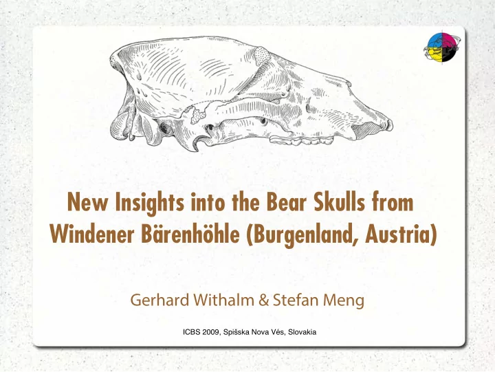
- New Insights into the Bear Skulls from
New Insights into the Bear Skulls from Windener Brenhhle - - PowerPoint PPT Presentation
New Insights into the Bear Skulls from Windener Brenhhle (Burgenland, Austria) Gerhard Withalm & Stefan Meng ICBS 2009, Spi ska Nova Vs, Slovakia The Windener Brenhhle (2911/1) ICBS 2009, Spi ska Nova Vs, Slovakia
ICBS 2009, Spišska Nova Vés, Slovakia
ICBS 2009, Spišska Nova Vés, Slovakia
ICBS 2009, Spišska Nova Vés, Slovakia
ICBS 2009, Spišska Nova Vés, Slovakia
ICBS 2009, Spišska Nova Vés, Slovakia
There are two bear skulls from Windener Bärenhöhle (Burgenland, Austria), which repeatedly occur in the relevant literature because of their interesting morphology. These skulls were excavated during the 30ies of the last century and were first described by Kurt EHRENBERG (1938) and firstly referred to – in doubt – as Ursus spelaeus in an article on the excavations in Windener Bärenhöhle. Based on their flat skull (missing glabella), their dentition and their supposed Late Pleistocene age he concluded, that these skulls must have belonged to a hybrid between cave- and brown bear or, less likely, to a not yet fully developed cave bear. KOBY (1944) rejected the hybridization-hypothesis of Ehrenberg. Later
their problems, published by SPAHNI in 1954. In this article he attributed these skulls to U. spelaeus, regardless of their morphology.
ICBS 2009, Spišska Nova Vés, Slovakia
Later on THENIUS (1956) reviewed the material and referred the skulls to Ursus arctos priscus Goldfuss, a member of the brown bear group, which was the state of knowledge until 2004. In the sense of THENIUS (1956) U. a. priscus is a big brown bear that coexisted with the cave bear group during Late Pleistocene, equipped with several characters that remind us on members of the cave bear
by its morphology as well as its ecological needs. There are also some similarities with brown bears from Asia, especially with U. a. piscator and
Ursus arctos priscus (GOLDFUSS) is possibly synonymous with U. taubachensis RODE and with U. a. nemoralis (DEGERBØL).
ICBS 2009, Spišska Nova Vés, Slovakia
To resolve the problem of these skulls, Prof. Rabeder decided to ask Michael Hofreiter if he would be interested in this subject and he agreed. In 2004 the skulls were analyzed by means of palaeo-DNA-analysis, which was carried out by Michael Hofreiter from the Max Planck Institute for Evolutionary Anthropology in Leipzig (Germany). The results of this work lead to the attribution of these skulls to a typical Ursus arctos, interestingly of the West-group. So the systematic position
ICBS 2009, Spišska Nova Vés, Slovakia
Definition 380 330 262 322 207 180 139 179 173 150 123 143 215 189 150 182 186 170 130 163 81 71 66 69 34 33 33 33 106 94 76 89 33 28 34 37 56 48 44 50 Distance Skull 1 Skull 2
Serbia, IPUW Bosnia, IPUW
Basilar length (prosthion – basion) Cranial length (basion – postdentale) Facial length (prosthion – postdentale) Palatal length (prosthion – staphylion) Width of mastoid Width of condyli occipitales Width of foramen magnum Occipital height (basion – acrocranium) Diastemal length Palatal width (behind staphylion, min.)
Abbreviation: IPUW – Intitute of Palaeontology, University of Vienna, data from THENIUS (1956:157)
ICBS 2009, Spišska Nova Vés, Slovakia
The specimens were scanned at the Department of Radiology of the Kaiser-Franz-Joseph-Spital, a hospital in the southern part of Vienna, with a dual source, multi-slice CT scanner (Somatom Definition, Siemens, Erlangen, Germany). The examinations were carried out during helical scanning (80 kVp, 160 mAseff) using a 20 x 0.6 mm detector configuration and a table feed of 8.4 mm per rotation. Axial sections were reconstructed every 0.3 mm with a section thickness of 0.6 mm. Three-dimensional post-processing was performed with the Osirix 3.5.1, 64 bit (Antoine Rosset, Joris Heuberger) software on a Mac Pro Dual Quad-Core workstation (Apple Inc.).
ICBS 2009, Spišska Nova Vés, Slovakia
Photo: Siemens
ICBS 2009, Spišska Nova Vés, Slovakia
Photo: Grey's Anatomy, p. 159
ICBS 2009, Spišska Nova Vés, Slovakia
Preparation+ Photo: Uwe Gille
ICBS 2009, Spišska Nova Vés, Slovakia
3D reconstruction in sagittal section of extant Hyaena hyaena. Scan: VU-Wien, Insitut für Bildgebende Diagnostik, Dr. Elisabeth Mayrhofer, RTA: S. Dengg
ICBS 2009, Spišska Nova Vés, Slovakia
ICBS 2009, Spišska Nova Vés, Slovakia
ICBS 2009, Spišska Nova Vés, Slovakia
ICBS 2009, Spišska Nova Vés, Slovakia
To warm the air for breathing. To insulate against cold or high temperatures. To make a more impressive voice (resonance). To extend the area for the insertion of M. masseter. To provide weight reduction for the skull. To provide a little of all of the aforementioned. To …
ICBS 2009, Spišska Nova Vés, Slovakia
There is an observable difference in the construction of
Brown bears usually do not have sinuses extending into
Cave bears usually have sinuses extending into the ossa
The relatively smallest sinuses can be found in Ursus
ICBS 2009, Spišska Nova Vés, Slovakia
ICBS 2009, Spišska Nova Vés, Slovakia
Sutura squamosa Sutura lambdoidea Sutura frontomaxillaris Meatus acusticus externus Sutura nasomaxillaris Sutura zygomaticomaxillaris Cellula sinus frontalis
ICBS 2009, Spišska Nova Vés, Slovakia
ICBS 2009, Spišska Nova Vés, Slovakia
ICBS 2009, Spišska Nova Vés, Slovakia
Sutura frontalis Sutura squamosa Sutura zygomaticomaxillaris Sutura nasomaxillaris Sutura lambdoidea Sutura frontomaxillaris Sutura coronalis Septum intersinuale frontale Apertura sinus frontalis
ICBS 2009, Spišska Nova Vés, Slovakia
ICBS 2009, Spišska Nova Vés, Slovakia
ex: THENIUS, 1956:Taf. 1
ICBS 2009, Spišska Nova Vés, Slovakia
ex: THENIUS, 1956:Taf. 1
ICBS 2009, Spišska Nova Vés, Slovakia
The skulls are flat, i.e. there is nothing like a glabella.
Only the skull of U. maritimus shows an even flatter
The occlusal surfaces of the teeth are more intensely
Their size is impressive.
ICBS 2009, Spišska Nova Vés, Slovakia
ICBS 2009, Spišska Nova Vés, Slovakia
ICBS 2009, Spišska Nova Vés, Slovakia
ICBS 2009, Spišska Nova Vés, Slovakia
ICBS 2009, Spišska Nova Vés, Slovakia
ICBS 2009, Spišska Nova Vés, Slovakia
ICBS 2009, Spišska Nova Vés, Slovakia
ICBS 2009, Spišska Nova Vés, Slovakia
ICBS 2009, Spišska Nova Vés, Slovakia
ICBS 2009, Spišska Nova Vés, Slovakia
We do not know enough about construction and
More information about morphology of sinuses should be
It's probably time for a cooperation with (veterinary)
ICBS 2009, Spišska Nova Vés, Slovakia