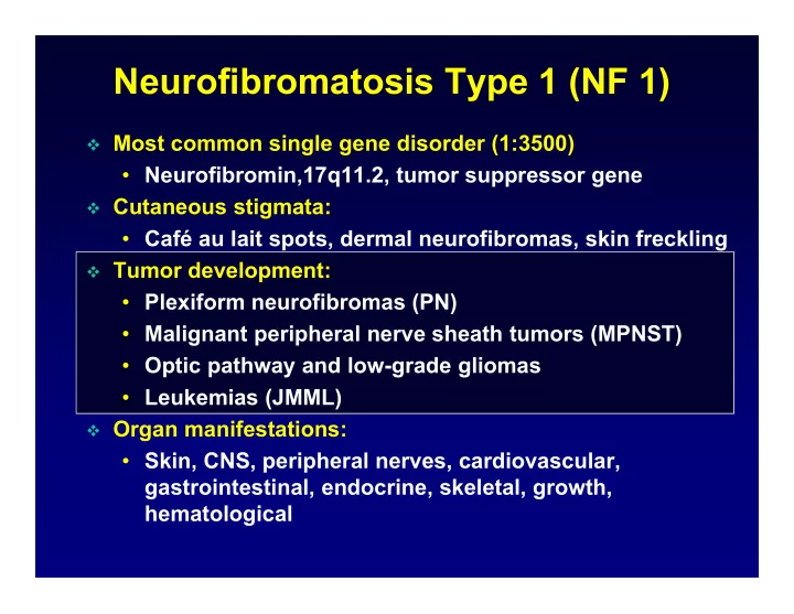

Neurofibromatosis Type 1 (NF 1) Most common single gene disorder (1:3500) • Neurofibromin,17q11.2, tumor suppressor gene Cutaneous stigmata: • Café au lait spots, dermal neurofibromas, skin freckling Tumor development: • Plexiform neurofibromas (PN) • Malignant peripheral nerve sheath tumors (MPNST) • Optic pathway and low-grade gliomas • Leukemias (JMML) Organ manifestations: • Skin, CNS, peripheral nerves, cardiovascular, gastrointestinal, endocrine, skeletal, growth, hematological
NF1 Tumor Development p120 GAP GTPase-Activating Proteins Neurofibromin GTP GDP Receptor Growth factor Targets for NF1 Tumors Ras pathway M-TOR Angiogenesis EGFR Kit, TGF- Mast cell
Plexiform Neurofibromas Involve multiple nerve fascicles/branches (25%) Congenital, erratic growth, large size and complex shape Disfigurement, functional impairment, life-threatening Malignant transformation to MPNST (8-13%) Surgical resection only standard treatment Medical treatment may reduce morbidity and prevent cancers
Trial Design Measure drug effect: Randomized Endpoints placebo-controlled Sensitively measure cross-over tumors: Vol. MRI Agents Access: CTEP, Pharma, FDA Response NF1 Trials Evaluation Infrastructure Centrally at NCI Collaborators Informal consortium, DoD NF1 consortium, Coop. groups (SARC) Referral Base Funding Nationwide Support for collaborators: DoD, Bench to Bedside
Clinical Drug Development Cancer NF1 Study Objective Endpoint Trials in NF1 Preclinical Start dose Toxicology in Chronic toxicity human trial animals Reproduction toxicity Phase I MTD Toxicity Chronic dosing PK Redefine DLT, MTD Phase II Activity Response Response unrealistic TTP Progression difficult to measure Unknown NH Phase III Efficacy Survival Near normal survival QOL QOL MTD, Maximum Tolerated Dose; DLT, Dose-Limiting Toxicity; TTP, Time To Progression; QOL, Quality Of Life; PK, Pharmacokinetics
Complex Plexiform Neurofibromas Measuring change in PN size difficult with standard criteria (WHO, RECIST)
Volumetric MRI Analysis: MEDx STIR Sequence Define Border Histogram Analysis Final Tumor Border Threshold Pixel number Volume 91 ml Pixel signal intensity
Growth Rate and Pattern of PNs 3 years old 30 30 Percent Change in PN Volume 5 years old 16 years old 25 25 Progressive Disease: 20 20 PN Volume 20% 15 15 10 10 21 years old 5 5 0 0 -5 -5 0 0 6 6 12 12 18 18 24 24 30 30 Time (months) Time (months)
Longitudinal Volumetric MRI of PN • 49 patients with 61 PN, median age 8.3 years (3.3-25) • Observation period 34 months (18-70 months) • PN volume at baseline 471 ml (31-5573 ml) PN Volume Body Weight % CHANGE IN BODY WEIGHT PER YEAR % CHANGE IN BODY WEIGHT PER YEAR % CHANGE IN PN VOLUME PER YEAR % CHANGE IN PN VOLUME PER YEAR y = 134.3 * e^(-0.31x) R 2 = 0.4 y = 134.3 * e^(-0.31x) R 2 = 0.4 60 60 60 60 40 40 40 40 20 20 20 20 0 0 0 0 -20 -20 -20 -20 0 0 5 5 10 10 15 15 20 20 25 25 30 30 0 0 5 5 10 10 15 15 20 20 25 25 30 30 AGE (YEARS) AGE (YEARS) AGE (YEARS) AGE (YEARS)
Conclusions Volume Analysis PN Volumetric MRI sensitively measures PN growth PN growth rate varies among patients, but is constant within a patient PNs grow more rapidly in younger patients Age stratification for treatment trials is indicated Body growth does not account for more rapid growth of PN in young children Drug development for PN should target young patients 11 mo. 17 mo. 25 mo. 36 mo. 11 mo. 17 mo. 25 mo. 36 mo.
Tipifarnib Farnesyltransferase Inhibitor Cl Cl NH 2 N N O N H 3 C CH 3 Mechanism of Action: Targets RAS farnesylation Route of Administration: Oral, twice daily for 21 days Toxicity Profile: Myelosuppression, rash, GI Cancer Development: Leukemias/MDS, breast
Pediatric Phase I Trial of Tipifarnib Eligibility: Age 2-18 years NF1 and solid tumors Endpoints: MTD, toxicities, PK, PD Schedule: Oral every 12 hours for 21 days followed by 7 days rest Dose levels: 150 (n=4), 200 (n=13) MTD, 275 (n=12), 375 (n=7) mg/m 2 /dose
Pediatric Tipifarnib Phase I Trial Characteristics NF1 Solid Tumor Patients entered (N) 21 25 Median (range) Age (yr) 7 (5-16) 15 (5-18) Diarrhea (1) Plt (3), ANC (2), Rash (1) DLT ANC (1), Rash (1) NV (1), FN (1) 2968 3570 ANC baseline (/µL) (1495-8520) (2330-7200) ANC nadir (% decrease) 32 (0-87) 37 (0-100) 819 680 CL/F (mL/min/m 2 ) (280-2070) (162-4310) Median (range) cycle # 10 (1-32) 1 (1-4) Cumulative toxicity None Not evaluable
Tipifarnib Pharmacokinetics (200 mg/m 2 ) 1000 C 12 h : 146 nM C 12h : 103 nM 100 Solid Tumor NF1 0 4 8 12 Hours • 70% inhibition of FTase in PBMCs
Phase II Trial of Tipifarnib for PN Double-blinded, placebo-controlled, flexible cross-over Endpoint: Time to progression (PN volume ≥ 20%) Phase A Phase B P P Tipifarnib Tipifarnib r r o o g g r r e e Randomize s s s s i i o o Placebo Placebo n n
Status of Tipifarnib Phase II Trial Patients: 58 (35 m, 23 f), median age: 8 years (3-21 yrs.) Off Study for: Enrolled on Phase A (n=58) Withdrew (n=3) Toxicity (n=2) MPNST (n=2) No Stable on P Non-Adherence (n=1) Phase A (n=22) Death (n=1) Yes Off Study for: Enrolled on Phase B (n=27) Withdrew (n=1) Toxicity (n=2) Non-Adherence (n=1) No Stable on P Phase B (n=11) Yes Off Study (n=12) P = Progression ( ≥ 20% volume increase)
Time to Progression (Phase A) 54 Patients Progression Free Survival 100 1D (20% ) 80 (25% ) 2D (20% ) 3D 3D 60 40 20 0 0 12 24 36 48 Months
Conclusions Tipifarnib Phase II Trial Tipifarnib / placebo toxicity indistinguishable Volumetric MRI analysis more sensitive than standard criteria in detecting progression Progression by volumetric MRI is a valid endpoint Randomized flexible cross-over design is feasible Placebo arm will serve as historical control group for other ongoing trials
Malignant Peripheral Nerve Sheath Tumor MPNST Characteristics Sporadic NF1 Incidence (%) 0.001 8-13 Age at diagnosis (yrs.) 40-62 26-36 Development De novo In PN Pain, rapid growth, neurologic Clinical findings compromise Molecular biology Not distinct Chemotherapy response % 55 18 5-year survival % 42-57 16-38
Phase II Trial of Neoadjuvant Chemotherapy in Sporadic and NF1 Associated High Grade Unresectable MPNST NF1 IA x 2 IE x 2 I - Ifosfamide Response Local MPNST Chemotherapy A - Adriamycin Evaluation Control E - Etoposide Sporadic IA x 2 IE x 2 PET PET Surgery 3D MRI 3D MRI MRI XRT Primary objective: • Response rate after 4 cycles of chemotherapy in NF1 and sporadic MPNST • Target response rate (CR or PR by WHO criteria): 40% Secondary objectives: • Response evaluation with 18 FDG-PET, 3-D MRI, pathology (% necrosis) • Molecular biology, tissue microarray, serum proteomics • Epidemiology of MPNST (NF1 vs. sporadic) A collaborative effort of NCI, SARC, and NF1 centers, funded through DoD grant
Future Directions in NF1 Collaborative natural history study (Trans-NIH): • Geno-, phenotyping, optic gliomas, hormonal influence, cognitive function Separate NF1 phase I trials (after cancer trials) Phase II PN trials within DoD NF1 Consortium FDG-PET for the diagnosis of MPNST within PN Phase II trials in recurrent MPNST within DoD NF1 Consortium Develop methods to measure dermal and spinal neurofibromas Clinical trials for dermal neurofibromas
FDG-PET Imaging of NF1 MPNST MPNST arising in Pelvic PN Neck PN
Future Directions in NF1 Collaborative natural history study (Trans-NIH): • Geno-, phenotyping, optic gliomas, hormonal influence, cognitive function Separate NF1 phase I trials (after cancer trials) Phase II PN trials within DoD NF1 Consortium FDG-PET for the diagnosis of MPNST within PN Phase II trials in recurrent MPNST within DoD NF1 Consortium Develop methods to measure dermal and spinal neurofibromas Clinical trials for dermal neurofibromas
Measurement of Dermal Neurofibromas Volume photography 3-D measurements of skin surface Dermal Neurofibroma Natural history and biology study of dermal neurofibromas NHGRI and NCI collaboration
Recommend
More recommend