
SLIDE 1
Neurocysticercosis in sub-Saharan Africa
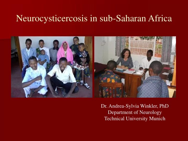
- Dr. Andrea-Sylvia Winkler, PhD
Department of Neurology Technical University Munich
Neurocysticercosis in sub-Saharan Africa Dr. Andrea-Sylvia Winkler, - - PowerPoint PPT Presentation
Neurocysticercosis in sub-Saharan Africa Dr. Andrea-Sylvia Winkler, PhD Department of Neurology Technical University Munich (Sero-)prevalence of cysticercosis (worldwide) Worldwide 50 million people with cysticercosis (WHO 2005) = most
Department of Neurology Technical University Munich
Worldwide 50 million people with cysticercosis (WHO 2005) = most frequent cerebral helminthosis
about 45% in Tanzania (rt-24h Ab-detecting ELISA).
USA.
cases/year (many cases not reported – no seroprevalence studies)
Druet-Cabanac 2005).
Winkler et al. 2009).
10 20 30 40 50 60 70 80
Äthiopien Nigeria Nigeria Elfenbeink. Senegal Tansania Tansania Burkina F: Tansania Mali Togo Zambia Uganda Togo Tansania Benin Mali Togo Kenya Togo Madagaskar Senegal Benin Liberia Benin Nigeria Kamerun Elfenbeink. Kamerun Elfenbeink.
Prävalenz pro 1000 0 5,2 74,4 11,2 13,2
Druet-Cabanac 2005).
Winkler et al. 2009).
Druet-Cabanac 2005).
Winkler et al. 2009).
Druet-Cabanac 2005).
Winkler et al. 2009).
focal signs or diffuse brain damage obvious yes no
6-25 years
6-25 years
focal signs or diffuse brain damage obvious yes no focal signs/ focal neurology
(progressive)
6-25 years
6-25 years diffuse brain damage (non-
progressive)
focal signs or diffuse brain damage obvious yes no focal signs/ focal neurology
(progressive)
6-25 years
6-25 years further clinical work-up EEG/CT necessary further clinical work-up only in selected cases diffuse brain damage (non-
progressive)
further clinical work-up only in selected cases
close follow-up necessary close follow-up necessary close follow-up not necessary focal signs or diffuse brain damage obvious yes no focal signs/ focal neurology
(progressive)
6-25 years
6-25 years further clinical work-up EEG/CT necessary further clinical work-up only in selected cases drug of choice: 1.CBZ 2.PHT drug of choice: 1.CBZ 2.PHT or PHB drug of choice: children: PHT adults: PHB diffuse brain damage (non-
progressive)
further clinical work-up only in selected cases
CT scan Epileptic seizures and epilepsy most likely due to NCC Antigen ELISA Positive CT suggestive of NCC Negative CT scan refer back to the system
Confirmed as NCC
CT scan Epileptic seizures and epilepsy most likely due to NCC Antigen ELISA Positive CT suggestive of NCC Negative CT scan refer back to the system Negative
Confirmed as NCC
Immunoblot Positive Negative
signs of increased intracranial pressure, vasculitis, compression of the brainstem, spine or optic nerve.
treatment may be required; in subarachnoid forms high doses of both drugs and long treatment.
mg/kg for one day!
into CNS)
(Praziquantel > Albendazole)
pressure with „sudden death“; Combination with steroids and control-CTs are essential!
Active Parenchymal neurocysticercosis Transitional Inactive
Antihelminthics Steroids AED
Steroids
AED
(Steroids)
AED
Active Parenchymal neurocysticercosis Transitional Inactive
Antihelminthics Steroids AED
Extraparenchymal neurocysticercosis
Steroids
AED (Steroids) AED
Ventricular Subarachnoid*
Neurosurgery
Antihelminthics Steroids (AED) Neurosurgery
Steroids
Antihelminthics
* The racemose NCC form is a malignant version of the subarachnoid form.
Active or transitional stage Initiate AED treatment – CT control in 3-6 months
Cysts not resolved with or without seizure recurrence Cysts resolved but seizure recurrence Cysts resolved an no seizure recurrence Maintain AED and perform control CT as above Maintain AED at least for another year Withdraw AED
Active or transitional stage Initiate AED treatment – CT control in 3-6 months
Cysts not resolved with or without seizure recurrence
Inactive (calcification) stage
Cysts resolved but seizure recurrence Cysts resolved an no seizure recurrence
Initiate AED treatment
Seizure recurrence No seizure recurrence for
Withdraw AED Maintain on AED until no seizure recurrence for one year Maintain AED and perform control CT as above Maintain AED at least for another year Withdraw AED
Carpio and Ross 2012 http://emedicine.medscape.com/article/1168784-overview#a0199
Performance of CT scan (not older than a couple
Epileptic seizures and epilepsy most likely due to NCC (narrow window between first seizure and first CT scan) CT scan possible – active NCC confirmed
Performance of CT scan (not older than a couple
Epileptic seizures and epilepsy most likely due to NCC (narrow window between first seizure and first CT scan) Praziquantel and steroids (encephalitis steroids only) CT scan possible – active NCC confirmed
Performance of CT scan (not older than a couple
Epileptic seizures and epilepsy most likely due to NCC (narrow window between first seizure and first CT scan) Praziquantel and steroids (encephalitis steroids only) Follow up with CT? CT scan possible – active NCC confirmed Follow up with serology? Treatment for how long? What to do with defaulters? What to do with treatment failure?
Performance of CT scan (not older than a couple
Epileptic seizures and epilepsy most likely due to NCC (narrow window between first seizure and first CT scan) Praziquantel and steroids (encephalitis steroids only) Follow up with CT? CT scan possible – active NCC confirmed CT scan not possible a) none available b) financial constraints Follow up with serology?
Treatment with steroids (without antihelminthics) based on serology under very close observation by a specialist?? Or Symptomatic treatment, i.e. AED only, and follow wait and see policy???
Treatment for how long? What to do with defaulters? What to do with treatment failure?
account for between 10% and 40% of all NCC cases (Carpio & Ross 2012 (medscape)).
80% due to calcifications.
2011).
(Ndimubanzi et al. 2010).
extraparenchymal forms.