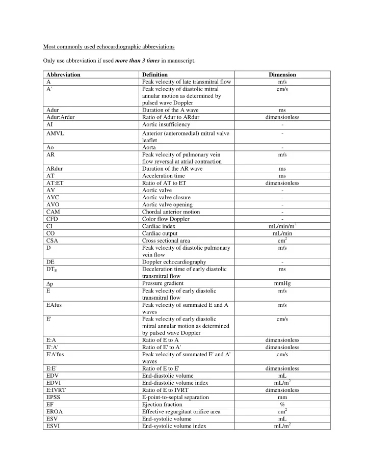

Most commonly used echocardiographic abbreviations Only use abbreviation if used more than 3 times in manuscript. Abbreviation Definition Dimension A Peak velocity of late transmitral flow m/s A' Peak velocity of diastolic mitral annular motion as determined by pulsed wave Doppler cm/s Adur Duration of the A wave ms Adur:Ardur Ratio of Adur to ARdur dimensionless AI Aortic insufficiency
- AMVL
Anterior (anteromedial) mitral valve leaflet
- Ao
Aorta
- AR
Peak velocity of pulmonary vein flow reversal at atrial contraction m/s ARdur Duration of the AR wave ms AT Acceleration time ms AT:ET Ratio of AT to ET dimensionless AV Aortic valve
- AVC
Aortic valve closure
- AVO
Aortic valve opening
- CAM
Chordal anterior motion
- CFD
Color flow Doppler
- CI
Cardiac index mL/min/m2 CO Cardiac output mL/min CSA Cross sectional area cm2 D Peak velocity of diastolic pulmonary vein flow m/s DE Doppler echocardiography
- DTE
Deceleration time of early diastolic transmitral flow ms p Pressure gradient mmHg E Peak velocity of early diastolic transmitral flow m/s EAfus Peak velocity of summated E and A waves m/s E' Peak velocity of early diastolic mitral annular motion as determined by pulsed wave Doppler cm/s E:A Ratio of E to A dimensionless E':A' Ratio of E' to A' dimensionless E'A'fus Peak velocity of summated E' and A' waves cm/s E:E' Ratio of E to E' dimensionless EDV End-diastolic volume mL EDVI End-diastolic volume index mL/m2 E:IVRT Ratio of E to IVRT dimensionless EPSS E-point-to-septal separation mm EF Ejection fraction % EROA Effective regurgitant orifice area cm2 ESV End-systolic volume mL ESVI End-systolic volume index mL/m2