Outline Echocardiographic Assessment of RV Conventional Doppler - - PowerPoint PPT Presentation

outline
SMART_READER_LITE
LIVE PREVIEW

Outline Echocardiographic Assessment of RV Conventional Doppler - - PowerPoint PPT Presentation

6/22/2013 Outline Echocardiographic Assessment of RV Conventional Doppler Function and Failure in Children with Pulmonary Hypertensive Vascular Disease 2-D assessment of RV function Annular motion Assessment of myocardial


slide-1
SLIDE 1

6/22/2013 1

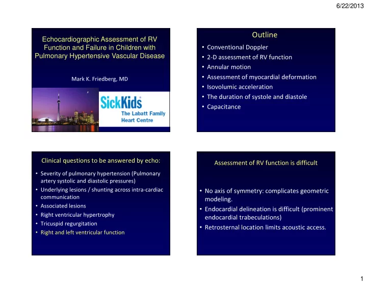
Echocardiographic Assessment of RV Function and Failure in Children with Pulmonary Hypertensive Vascular Disease

Mark K. Friedberg, MD

Outline

  • Conventional Doppler
  • 2-D assessment of RV function
  • Annular motion
  • Assessment of myocardial deformation
  • Isovolumic acceleration
  • The duration of systole and diastole
  • Capacitance
  • Severity of pulmonary hypertension (Pulmonary

artery systolic and diastolic pressures)

  • Underlying lesions / shunting across intra-cardiac

communication

  • Associated lesions
  • Right ventricular hypertrophy
  • Tricuspid regurgitation
  • Right and left ventricular function

Clinical questions to be answered by echo: Assessment of RV function is difficult

  • No axis of symmetry: complicates geometric

modeling.

  • Endocardial delineation is difficult (prominent

endocardial trabeculations)

  • Retrosternal location limits acoustic access.
slide-2
SLIDE 2

6/22/2013 2

RV structure

Chin, Coronary Artery Disease 2005, 16:13–18 Anderson, Semin Thorac Cardiovasc Surg Pediatr Card Surg Ann 2007 10:76

RV Free Wall IVS

Which is worse?

‘Eyeball’ assessment still most common method

RV does not tolerate acute increases in afterload

MacNee W. Am J Respir Crit Care Med. 1994;150: 833–852.

slide-3
SLIDE 3

6/22/2013 3

Adaptation to increased afterload

15 year old girl with presenting with pulmonary emboli RV dilatation as initial response to increased afterload

Courtesy Luc Mertens

RV dimensions

Lopez, J Am Soc Echocardiogr 2010;23:465-95

Parameters of longitudinal function

Tricuspid annular plane systolic excursion (TAPSE)

Koestenberger, JASE 2009

slide-4
SLIDE 4

6/22/2013 4

TAPSE correlates with RV EF

López-Candales, Am J Cardiol 2006;98:973 Koestenberger, Congenit Heart Dis. 2012;7:250

grey diamonds PAH-CHD patients black triangles TOF patients

Deviation of TAPSE values in patients with PAH-CHD and TOF from mean age- related reference values is shown (TAPSE D ).

Deviation of TAPSE values from mean age- related reference values

TAPSE correlates with stroke volume

Forfia, Am J Respir Crit Care Med 2006 174:1034–1041

Ability of TAPSE to predict RV indexed stroke volume of 29 ml/ m2. Forfia, Am J Respir Crit Care Med, 2006 174:1034

TAPSE correlates with survival

Tricuspid tissue Doppler velocities

Eidem, J Am Soc Echocardiogr 2004;17:212-21

slide-5
SLIDE 5

6/22/2013 5

TDI severe PAH

Tissue Displacement

Pre-tricuspid Eisenmenger

Normal TAPSE Low FAC% due to apical dysfunction

13y, iPAH, PAH worsening, pneumonia

slide-6
SLIDE 6

6/22/2013 6 Assessment of regional RV function by strain

Dambrauskaite, J Am Soc Echocardiogr 2007;20:1172

Fractional area of change

Mertens, L. L. & Friedberg, M. K. Nat. Rev. Cardiol, 2010

3-D assessment of RV volumes and EF

LV function influences TAPSE

López-Candales, AJC 2006;98:973

slide-7
SLIDE 7

6/22/2013 7

Right atrial size

Bustamante-Labarta, JASE 2002;15:1160

Pericardial effusion

Douglas, Circulation 1989;80:353

Other important 2-D parameters

Conventional echo parameters in children with PAH

Kassem E, Am Heart J. 2013;165:1024-31

Conventional echo parameters in children with PAH

Kassem E, Am Heart J. 2013;165:1024-31

iPAH All

Assessment of myocardial function

slide-8
SLIDE 8

6/22/2013 8

Strain and strain rate

Strain correlates with stroke volume

Urheim, AJC 2005;96:1173

*SV determined at cath

RV myocardial function

Urheim, AJC 2005;96:1173

Isovolumic acceleration is a relatively load independent measure of RV function

Vogel, Circulation. 2002;105:1693-1699

slide-9
SLIDE 9

6/22/2013 9

IVA is relatively load independent

Urheim, AJC 2005;96:1173

RV strain in pediatric iPAH patients

Submitted

RV strain in pediatric iPAH patients

Submitted

Back to Doppler….

slide-10
SLIDE 10

6/22/2013 10

Doppler hemodynamics

+ Rap …

MPI for RV function

“ “ “ “b” ” ” ” is RV ejection time “ “ “ “a” ” ” ” is duration of tricuspid valve regurgitation

Eidem , Am J Cardiol 2000;86:654 Mertens, L. L. & Friedberg, M. K. Nat. Rev. Cardiol, 2010

Problems with MPI

  • Cannot assess RV inflow and outflow simultaneously
  • HR has to be similar in both measurements
  • ICT is short in the normal RV -> IRT becomes more

important (itself an independent index)

  • Measurement of small intervals-prone to

measurement error

The S/D Duration ratio

Friedberg, JASE, 2006;19:1326

slide-11
SLIDE 11

6/22/2013 11

SD ratio in the RV in HLHS

Friedberg, J Am Soc Echocardiogr 2007;20:749

SD ratio in PAH

Alkon, Am J Cardiol 2010;106:430 Gan . Am J Physiol 2006; 290:1528

Ventricular interactions in PAH

Alkon, Am J Cardiol 2010;106:430

The S/D duration ratio in PAH

Marcus, J Am Coll Cardiol 2008; 51:750

slide-12
SLIDE 12

6/22/2013 12

S/D ratio=2.3 S/D ratio=1.3

13y, iPAH, pneumonia, PAH worsening

Echo predictors of mortality in Eisenmenger’s

Moceri, Circulation. 2012;126:1461-1468

Ventricular-vascular interactions

J Am Soc Echocardio gr 2006; 19:1045

slide-13
SLIDE 13

6/22/2013 13

Pulmonary arterial capacitance

PAC = RV stroke volume/pulmonary artery pulse pressure

Sajan, Am Heart J 2011;162:562

Pulmonary arterial capacitance by echo

Friedberg, J AmSoc Echocardiogr 2006;19: 559 Friedberg, J Am Soc Echocardiogr 2007;20:186

RV stroke work

DiMaria, ASE 2013

Summary

  • Assessment of RV function is important in PAH.
  • Echo is the mainstay of imaging in PAH, but has important

limitations.

  • Newer methods can be combined with conventional

assessment to provide more comprehensive evaluation.

  • Further validation of the newer methods in clinical practice

is needed.

  • Left ventricular function and ventricular-vascular

interactions should be accounted for

slide-14
SLIDE 14

6/22/2013 14

Thank you

  • Can be performed relatively safely and is usually well

tolerated (even when baseline oxygen saturation levels are < 80%.

  • May be useful for detecting atrial septal defects / patent

ductus arteriosus and for imaging pulmonary veins.

  • Should be performed in lung transplant candidates

(detect unsuspected intracardiac defects/ shunts, proximal pulmonary artery thrombus).

  • Gorcsan. Transesophageal echocardiography to evaluate patients with severe

pulmonary hypertension for lung transplantation. Ann Thorac Surg. 1995; 59:717-22.

Transesophageal echocardiography

The Evolution of Echocardiography…………

A-Mode……M-mode……2D…Doppler…Color- flow Doppler……COMPUTERS!!!!!!!!

slide-15
SLIDE 15

6/22/2013 15 Acoustic Quantification Echocardiography

Geva, Circulation. 1998;98:339

Assessing Myocardial Deformation

Courtesy of Piet Claus

RV strain by speckle tracking The S/D ratio: Dilated Cardiomyopathy

Friedberg, Am J Cardiol 2006;97:101–105

slide-16
SLIDE 16

6/22/2013 16

SD ratio in PHTN

  • higher systolic, diastolic and mean PAP
  • higher RVSP, RV/BP ratio
  • higher PVR after exposure to 40ppm NO
  • lower RV FAC%, lower RVEF (MRI)
  • higher RVED
  • lower systemic blood pressure
  • shorter 6 minute walk distance
  • younger age at diagnosis
  • non-repaired CHD

A higher S/D ratio is associated with:

3D assessment of RV function

Marcus, J Am Soc Echocardiogr 2002;15: 1145

What are we trying to capture? RV VVI

Kutty Echocardiography, Vol 25, 2008

slide-17
SLIDE 17

6/22/2013 17

A smaller TAPSE may originate from RV mechanical delay

Lopez-Candales Am J Cardiol 2005;96:602

Nelson Schiller and Annular excursion

Isaaz K, Munoz del Romeral L, Lee E, Schiller NB. Quantitation of the motion of the cardiac base in normal subjects by Doppler

  • echocardiography. J Am Soc Echocardiogr. 1993 Mar-Apr;6(2):166-76.

“Because the motion of the base of the heart plays a central role in its filling and emptying, we developed an original method to characterize the base motion dynamics throughout each cycle by use of pulsed Doppler echocardiography. A 100 Hz wall filter and low gain settings were used to record the low-frequency, high- energy Doppler signals generated by the motion of the base. From the apical four-chamber view, the sample volume was placed at the lateral margin and at the common septal margin of the tricuspid and mitral annuli.” “The method reported in the present study allows a more informative noninvasive quantitation of the cardiac base motion derived from measurements of its velocity, excursion, and acceleration.”

What should we measure?

Celermajer and Marwick, Int J Card (2008) 125 :294–303

M-mode

slide-18
SLIDE 18

6/22/2013 18

RV strain is influenced by afterload

Weidemann, Ped Cardiol, 2002;23:292

Normal RV TDI/ strain values in children

Mertens and Friedberg Nature Reviews Cardiology. (2010): p551.

TAPSE correlates with RV EF

López-Candales, Am J Cardiol 2006;98:973 Lissin, Am J Cardiol 2004;93:654–657

TGA after ASO with PHT

TAPSE NORMAL FOR AGE Tissue Doppler

slide-19
SLIDE 19

6/22/2013 19

REDUCED APICAL FUNCTION

TGA after ASO with PHT Improved LV filling after relief of RV outflow

  • bstruction

Lurz, EHJ 2009 30, 2266–2274

Improved LV filling after relief of RV outflow

  • bstruction

Lurz, EHJ 2009 30, 2266–2274