Monitoring of nanoparticles uptake by biological cells via far-field - - PowerPoint PPT Presentation

monitoring of nanoparticles uptake by biological cells
SMART_READER_LITE
LIVE PREVIEW

Monitoring of nanoparticles uptake by biological cells via far-field - - PowerPoint PPT Presentation

Monitoring of nanoparticles uptake by biological cells via far-field spectroscopy of localized surface plasmon resonance Dadadzhanov Daler 1, 2 , Vartanyan Tigran 1 , and Alina Karabchevsky 2 1 ITMO University, 49 Kronverksky Ave., 197101, St.


slide-1
SLIDE 1

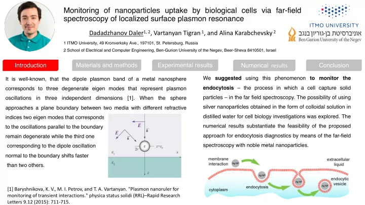
It is well-known, that the dipole plasmon band of a metal nanosphere corresponds to three degenerate eigen modes that represent plasmon

  • scillations

in three independent dimensions [1]. When the sphere approaches a plane boundary between two media with different refractive indices two eigen modes that corresponds to the oscillations parallel to the boundary remain degenerate while the third one corresponding to the dipole oscillation normal to the boundary shifts faster than two others. We suggested using this phenomenon to monitor the endocytosis – the process in which a cell capture solid particles – in the far field spectroscopy. The possibility of using silver nanoparticles obtained in the form of colloidal solution in distilled water for cell biology investigations was explored. The numerical results substantiate the feasibility of the proposed approach for endocytosis diagnostics by means of the far-field spectroscopy with noble metal nanoparticles. [1] Baryshnikova, K. V., M. I. Petrov, and T. A. Vartanyan. "Plasmon nanoruler for monitoring of transient interactions." physica status solidi (RRL)–Rapid Research Letters 9.12 (2015): 711-715.

Dadadzhanov Daler1, 2, Vartanyan Tigran 1, and Alina Karabchevsky 2

Monitoring of nanoparticles uptake by biological cells via far-field spectroscopy of localized surface plasmon resonance

1 ITMO University, 49 Kronverksky Ave., 197101, St. Petersburg, Russia 2 School of Electrical and Computer Engineering, Ben-Gurion University of the Negev, Beer-Sheva 8410501, Israel

Introduction Materials and methods Experimental results Numerical results Conclusion

slide-2
SLIDE 2

Dadadzhanov Daler1, 2, Vartanyan Tigran 1, and Alina Karabchevsky 2

Monitoring of nanoparticles uptake by biological cells via far-field spectroscopy of localized surface plasmon resonance

1 ITMO University, 49 Kronverksky Ave., 197101, St. Petersburg, Russia 2 School of Electrical and Computer Engineering, Ben-Gurion University of the Negev, Beer-Sheva 8410501, Israel

Nd:YAG laser at the wavelength of 532 nm

Fabrication of silver nanoparticles by laser ablation

To find out the dependence of the plasmon resonance splitting contrast on the particle position for various optical properties of the adjacent media, we examined plasmon resonance excitation in a spherical nanoparticle with polarization in - x (top excitation) and -z (side excitation) direction. Herein, we assume that the biological medium has the refractive index 1.4, while the culture medium has 1.33.

The scheme of numerical model(

Introduction Materials and methods Experimental results Numerical results Conclusion

slide-3
SLIDE 3

300 400 500 600 700 800 0.0 0.2 0.4 0.6 0.8 1.0 1.2 1.4 1.6 1.8 2.0 2.2 2.4

Optical density[arb. u.] Wavelength [nm] 3 min 5 min 10 min 15 min 35 min 60 min §= 397 nm

a b

a b

80

in in in in

2000 4000 6000 8000 10000 12000 0.5 1.0 1.5 2.0 2.5

Number of pulse

  • Max. optical density [arb.u.]

40 60 80 100 120 140 160

FWHM [nm]

b

Number of particles P a r t i c l e s i z e [ n m ] 10 20 40 60 80 40 30 20 50 60 70

b

300 350 400 450 500 550 600 0.0 0.1 0.2 0.3 0.4 0.5

Dlshift ~13 nm Optical density Wavelength [nm]

Ag NPs Ag NPs with BSA

To simulate the nanoparticle transition from subcutaneous water to cellular membrane, we measured the shift of the plasmon band maximum when the nanoparticles were transferred from water with a refractive index of 1.33 to a bovine serum albumin solution with a refractive index of 1.4.

Dadadzhanov Daler1, 2, Vartanyan Tigran 1, and Alina Karabchevsky 2

Monitoring of nanoparticles uptake by biological cells via far-field spectroscopy of localized surface plasmon resonance

1 ITMO University, 49 Kronverksky Ave., 197101, St. Petersburg, Russia 2 School of Electrical and Computer Engineering, Ben-Gurion University of the Negev, Beer-Sheva 8410501, Israel

Introduction Materials and methods Experimental results Numerical results Conclusion

slide-4
SLIDE 4
  • 100
  • 80
  • 60
  • 40
  • 20

20 40 60 80 100 398 399 400 401 402 403 404 405 406

LSPR max [nm] Position along z [nm] Top excitation Side excitation NP Cell

  • 100
  • 80
  • 60
  • 40
  • 20

20 40 60 80 100

  • 1.5
  • 1.0
  • 0.5

0.0 0.5 1.0 1.5

Splitting contrast [nm] Position alongz [nm] without membrane with membrane

  • 100 -80
  • 60
  • 40
  • 20

20 40 60 80 10 398 400 402 404 406 408 410

LSPR max [nm] Position along z [nm] Top excitation Side excitation NP Cell

With cell membrane Without cell membrane Dadadzhanov Daler1, 2, Vartanyan Tigran 1, and Alina Karabchevsky 2

Monitoring of nanoparticles uptake by biological cells via far-field spectroscopy of localized surface plasmon resonance

1 ITMO University, 49 Kronverksky Ave., 197101, St. Petersburg, Russia 2 School of Electrical and Computer Engineering, Ben-Gurion University of the Negev, Beer-Sheva 8410501, Israel

Introduction Materials and methods Experimental results Numerical results Conclusion

slide-5
SLIDE 5
  • As a result, a colloidal solution of silver NPs in water with diameters of 20 to 50 nm, free from stabilizing agents, was
  • btained
  • The possibility of using silver NPs obtained by laser ablation in water for monitoring the process of endocytosis was

confirmed

  • The theoretical results of the endocytosis model for single nanoparticle penetrated through a thin cell membrane has been

numerically developed

  • When the spherical nanoparticle approaches a cell, its triple degenerate dipole plasmon resonance splits into two

components: a non-degenerate mode, corresponding to oscillation along the line connecting the particle to the cell, and a doubly degenerate mode, corresponding to oscillation in perpendicular directions.

Contact facebook: https://www.facebook.com/principfree Email: daler.dadadzhanov@gmail.com Dadadzhanov Daler1, 2, Vartanyan Tigran 1, and Alina Karabchevsky 2

Monitoring of nanoparticles uptake by biological cells via far-field spectroscopy of localized surface plasmon resonance

1 ITMO University, 49 Kronverksky Ave., 197101, St. Petersburg, Russia 2 School of Electrical and Computer Engineering, Ben-Gurion University of the Negev, Beer-Sheva 8410501, Israel

Introduction Materials and methods Experimental results Numerical results Conclusion