SLIDE 1
(Please activate your clickers)
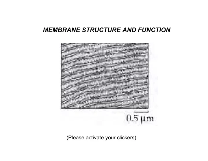
MEMBRANE STRUCTURE AND FUNCTION
SLIDE 2
Membrane structure
Lipid bilayer: hydrophobic fatty acid interior Phosphate + hydrophilic group exterior
SLIDE 3
Membrane structure
Proteins incorporated into the bilayer Transmembrane alpha-helix Hydrophobic amino acid side chains in the bylayer Hydrophilic amino acid side chains on the surface Extracellular carbohydrates “Fluid mosaic model” Often, “islands” of protein complexes
SLIDE 4
SLIDE 5 Membrane Functions
- 1. Platform for biochemical reactions
Enzymes and other organelles (e.g. ribosomes, microfilaments) attach to surface
SLIDE 6 Membrane Functions
Receptors bind to "ligand" (signal molecule) and transmit a signal across the membrane
SLIDE 7 Membrane Functions
Receptors on different cells bind them together
SLIDE 8 Membrane Functions
Receptors on different cells bind them together
SLIDE 9 Membrane Functions
- 4. Differential permeability
Lipid bilayer forms a barrier to diffusion. Channels, carriers, and pumps transport substances across membranes Allows cells to adjust pH and salt concentrations Allows cells to keep functional chemicals inside Allows cells to take up food molecules and exclude toxins Allows cells to separate chemical reactions that
- therwise would interfere with one another
Allows cells to store energy in a useful way
SLIDE 10 How molecules cross membranes
- 1. Dissolving in lipid layer
Small non-polar molecules (benzene, ethanol, O2, CO2) Works for artificial lipid bilayers (i.e., no proteins)
Small polar molecules (H2O, NH3) Also works for lipid bylayers Postulate transient pores
- 3. Channels, Carriers, Pumps
Specific materials transported Specific membranes Regulated in time and sometimes direction Proteins
SLIDE 11
Channels
Protein complex forming controlled hole for rapid flow "Downhill” (along energy gradient) “Gated” (opens, closes in response to stimulus) Channels known for Na+, K+, Cl-, Ca2+, and others, including H2O (aquaporins)!
SLIDE 12 Carriers
Proteins that shuttle molecules across a membrane: Operation: Carrier binds molecule—needs specific, reversible binding site. Carrier shifts to expose molecule to other side of membrane—needs flexibility. Carrier releases molecule. Empty carrier shifts to return binding site to original side
SLIDE 13
Why carriers are proteins:
Binding sites must be specific, reversible Proteins have multitude of possible shapes, it is possible to have a shape with a crevass to fit a particular transported molecule Variety of protein side chains provides variety of weak bonds (H-, electrostatic, hydrophobic, as well as Van der Waals); weak bonds give tight, but reversible bonds Flexibility of proteins allows shape changes— binding site can be accessible to either side
SLIDE 14
Pumps
Proteins that use energy to move molecules across a membrane Pumps known for H+, Ca2+, and Na+/K+, Mg2+, K+, K+/H+, P-lipid, heavy metals
SLIDE 15 How fungi take up and accumulate sugar (glucose) and K+
- 1. Proton pump in plasma membrane pumps H+ out
- f cell, forming gradients of [H+] and electric charge.
- 2. Carrier binds glucose and proton outside,
transports both to the inside; K+ enters the cell through open channels
- 4. Energy of H+ and charge gradients used to power
accumulation of glucose and K+ from low concentration solution.
SLIDE 16
How animal cells take up and accumulate K+ and glucose
Na+/K+ pump in plasma membrane replaces the H+ pump and forms Na+, K+, and electrical gradients.
SLIDE 17
In nerve cells that relay painful sensations in the body's tissues to the central nervous system, SCN9A encodes instructions for sodium channels that help the cells fire. In two of the disorders, people carry faulty versions of the gene and suffer crippling pain because their sodium channels open too easily or can't close. In the third disorder, which leaves patients unable to feel pain at all, SCN9A produces a protein that can't function.
"We wondered if more common, apparently harmless [changes] in the gene might give rise to an altered degree of pain threshold," says Geoffrey Woods, a medical geneticist at Cambridge University in the U.K., who discovered the genetic reason for this third disorder. One [genetic variant], found in 10% of the subjects, caused the greatest increase in reported pain between those who had it and those who didn't. When the team applied heat stimuli to 186 healthy women, they found that those with the rare version were more likely to have lower pain thresholds. It was as if the normal subjects had taken an ibuprofen, but the subjects with the rare SNP hadn't.