MOL2NET, 2017 , 3, doi:10.3390/mol2net-03-xxxx 2 25923, Pseudomonas - - PDF document

mol2net 2017 3 doi 10 3390 mol2net 03 xxxx 2 25923
SMART_READER_LITE
LIVE PREVIEW

MOL2NET, 2017 , 3, doi:10.3390/mol2net-03-xxxx 2 25923, Pseudomonas - - PDF document

MOL2NET, 2017 , 3, doi:10.3390/mol2net-03-xxxx 1 MOL2NET, International Conference Series on Multidisciplinary Sciences MDPI http://sciforum.net/conference/mol2net-03 Standardization of the Safety Level of the Use of DMSO in Viability Assays in


slide-1
SLIDE 1

MOL2NET, 2017, 3, doi:10.3390/mol2net-03-xxxx 1

MDPI

MOL2NET, International Conference Series on Multidisciplinary Sciences http://sciforum.net/conference/mol2net-03

Standardization of the Safety Level of the Use of DMSO in Viability Assays in Bacterial Cells

Raquel Carlos de Brito (E-mail: quelbrito1987@gmail.com)a, Gildoberg Nunes da Silva (E-mail: bergnunes22@gmail.com)a, Ticiane Costa Farias (E-mail: ticiane_92@hotmail.com)a, Paula Benvindo Ferreira (E-mail: paulabenvindo92@hotmail.com)b, Sávio Benvindo Ferreira (E-mail: saviobenvindo@gmail.com)c.

a Graduate Student, Center for Teacher Training (CFP), Federal University of Campina Grande

(UFCG), Cajazeiras campus, Paraíba, Brazil.

b Master, Postgraduate Program in Natural and Synthetic Bioactive Products, Federal University of

Paraíba, João Pessoa, Paraíba, Brazil.

c Substitute Professor of Nursing Academic Unit, Center for Teacher Training (CFP), Federal

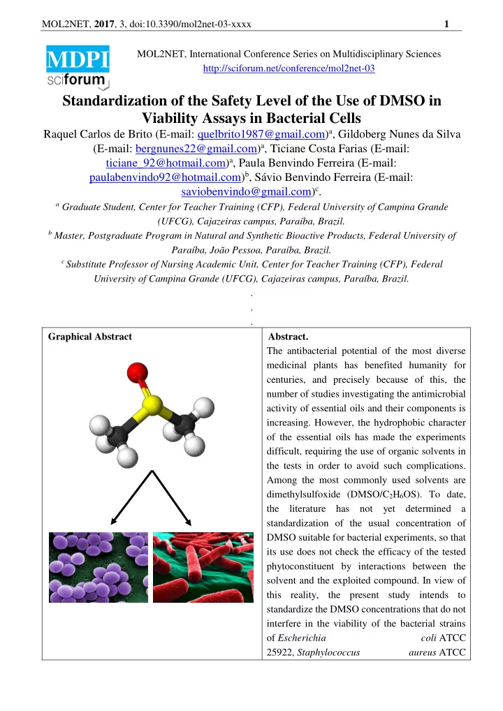
University of Campina Grande (UFCG), Cajazeiras campus, Paraíba, Brazil. . . . Graphical Abstract Abstract. The antibacterial potential of the most diverse medicinal plants has benefited humanity for centuries, and precisely because of this, the number of studies investigating the antimicrobial activity of essential oils and their components is

  • increasing. However, the hydrophobic character
  • f the essential oils has made the experiments

difficult, requiring the use of organic solvents in the tests in order to avoid such complications. Among the most commonly used solvents are dimethylsulfoxide (DMSO/C2H6OS). To date, the literature has not yet determined a standardization of the usual concentration of DMSO suitable for bacterial experiments, so that its use does not check the efficacy of the tested phytoconstituent by interactions between the solvent and the exploited compound. In view of this reality, the present study intends to standardize the DMSO concentrations that do not interfere in the viability of the bacterial strains

  • f Escherichia

coli ATCC 25922, Staphylococcus aureus ATCC

slide-2
SLIDE 2

MOL2NET, 2017, 3, doi:10.3390/mol2net-03-xxxx 2 25923, Pseudomonas aeruginosa ATCC 27853, Proteus mirabilis ATCC 25922 and Enterococcus faecalis ATCC 29212. In order to reach this objective, the disc diffusion method in Müller Hinton Agar of a reagent impregnated

  • n a filter paper disc was used, using different

concentrations of DMSO to determine the minimum inhibitory concentration (MIC) of the five microorganisms listed. The experiment was carried out in triplicate in order to safeguard the effectiveness

  • f the method tested. The

incubation was done in an oven at 35º C, for a period of 24 hours. For the test method the conclusions of the tests performed were expressed by the arithmetic mean of the diameter

  • f the inhibition halos formed around the disks.

The test demonstrates that for the concentrations and microorganisms tested, DMSO provides safety in its use. Introduction In the scientific field, extracts and essential oils from plants are used as natural sources of new compounds to combat bacterial infections. However, the estimation of the antibacterial activities of many plant-derived compounds is hampered by the low solubility of these compounds in water. Solubilizers, such as surfactants and solvents, among them dimethylsulfoxide (DMSO), have been used to solve this problem, but it may be difficult to distinguish the contribution in the antimicrobial activity of the solubilizer from the compounds under investigation [1]. DMSO was originally synthesized by Zaytsev in 1866, and has since been extensively investigated for possible industrial and biological utility, and a considerable amount of literature has developed on its properties and uses [2]. It is an organosulfur of formula C2H6OS of molecular weight 78 g/mol, boiling point 189 °C and freezing temperature 18.5 °C; an amphipathic molecule composed

  • f a polar domain characterized by sulfinyl and two non-polar methyl groups, which makes it capable of

solubilizing polar and non-polar substances and transposing hydrophobic barriers (Figure 1) [3]. These properties are important for pharmacological compounds that act as vehicles intracellularly as they allow non-water-soluble therapeutic and toxic agents to be generally soluble in DMSO [4]. Figure 1: Polarized form of dimethylsulfoxide

slide-3
SLIDE 3

MOL2NET, 2017, 3, doi:10.3390/mol2net-03-xxxx 3 An important biological action observed in DMSO is its ability to cross cell membranes; other pharmacological properties of DMSO include: immunomodulation; vasodilation; anti-platelet aggregation with protection against ischemic injury; diuretic, among others. The higher its concentration in water, the greater the penetrating capacity, the ideal range being between 70-90% of DMSO [5]. Thus, DMSO has been used as a commercial solvent since 1953, also possessing several other applications: therapeutic use, excipient for formulations in veterinary therapeutics, as a control group for testing natural products, for treating cells cultured in certain experiments and several in vitro studies. Despite the multiple applications of DMSO, its physiological and pharmacological characteristics as well as their effects are not fully understood, and further investigations into its pharmacological activities and which concentration of use is safe are required [6]. It has recently been found experimentally that DMSO has the ability to protect Escherichia coli from antimicrobial-mediated rapid death, interfering with the efficacy of the drugs tested, in this experiment DMSO inhibited death by ampicillin, kanamycin and two quinolones but had little effect on

  • MIC. DMSO-mediated protection correlated with ROS reduction [7]. Another assay also reported a

possible inhibition of microbial growth by interference of this solvent, thus, one can’t rule out the possibility that DMSO interfered in the experiment, possibly causing a potentiating effect of the antimicrobial activity of tested compounds [8]. In view of such reports, caution is advised in using DMSO-dissolved antimicrobials for short-term death assays as well as accurate investigations to find

  • ut which DMSO concentration is most reliable for use in such experiments.

Materials and Methods The microorganisms used for testing were Gram positive bacterial strains: Staphylococcus aureus ATCC 25923 and Enterococcus faecalis ATCC 29212; and Gram negative strains: Escherichia coli ATCC 25922, Pseudomonas aeruginosa ATCC 27853 and Proteus mirabilis ATCC 25922. Disk diffusion tests with different concentrations of dimethylsulfoxide (DMSO) were carried out in order to identify concentrations of DMSO capable of carrying out an antibacterial activity in the five

  • microorganisms. To test all concentrations, three petri dishes were used for each strain tested. The

incubation was done in an oven at 35º C, for a period of 24 hours. A suspension of the five microorganisms was performed, whose turbidity degree was 0.5 McFarland scale, corresponding to 1 x 10⁸ CFU/mL, which was spread with the aid of a Drigalsky loop. After sowing on the plate, each disk was impregnated with different concentrations of the reactants and pressed against the plate in order to ensure complete contact with the agar surface, being applied individually and evenly distributed, so that the distance from the center of the edge of the board did not exceed 24 mm. The plates containing Müller-Hinton Agar were inverted and placed in an oven at 35 ºC for 24 hours after the application of the discs therein, and thereafter the halos were read. The tests were performed and their results were expressed in mm by the arithmetic mean of the diameter of the inhibition halos formed around the discs during the disk diffusion test. Results and Discussion The disc diffusion method is performed to determine patterns of susceptibility to antibiotics according to the guidelines of the Institute of Clinical and Laboratory Standards (CLSI) [9]. It is an easy- to-perform and economical technique developed by the European Committee for Antimicrobial Susceptibility Testing (EUCAST), its standardized methods and clinical breakpoints have been adopted by clinical microbiology laboratories in Europe as well as in other parts of the globe [10].

slide-4
SLIDE 4

MOL2NET, 2017, 3, doi:10.3390/mol2net-03-xxxx 4 From the evaluation of the results recorded in Table 1, it was verified that there was no inhibition halo formation at any of the concentrations tested, that is, there was no impediment to the growth of the investigated microorganisms. This demonstrates that the use of DMSO as a chemical solvent in experiments with the probed strains, in concentrations not exceeding 80%, according to the assay performed, is considered safe, not causing any inhibition of microbial growth. Table 1: Verification of the antimicrobial activity of DMSO in different concentrations in the fusion disc method. Microorganism Substances / Halo (mm) AMC DMSO 80% 40% 20% 10% 5% 2,5% 1,75% 0,88%

  • S. aureus ATCC 25923

30 Ø Ø Ø Ø Ø Ø Ø Ø

  • E. faecalis ATCC 29212

22 Ø Ø Ø Ø Ø Ø Ø Ø

  • E. coli ATCC 25922

29 Ø Ø Ø Ø Ø Ø Ø Ø

  • P. mirabilis ATCC 25933

22 Ø Ø Ø Ø Ø Ø Ø Ø

  • P. aeruginosa ATCC 27853

33 Ø Ø Ø Ø Ø Ø Ø Ø AMC: amikacin disc (30 μg), Ø: absence of inhibition halo of bacterial growth. The reading of the results was done by measuring the diameter of the halos, in mm, formed around the discs containing the reagents, when greater than 6 mm it becomes visible indicating susceptibility of the microorganism to the substance tested [11]. The experiment was carried out by Filipe et al. [12], where the antimicrobial activity of the leaves

  • f Cydonia oblonga miller was tested against several microorganisms, including E. coli and S. aureus.

DMSO at 20% concentration was used in the assay and, corroborating our finding, the experiment did not detect any interference with the growth of these bacteria at the concentration used. Macieira [13] conducted a research on the antimicrobial activity of marine cyanobacteria, where eight bacterial species were selected: four Gram-positive (Staphylococcus aureus, Enterococcus faecalis, Enterococcus faecium and Bacillus subtilis) and four Gram-negative strains (Klebsiella pneumoniae, Escherichia coli, Pseudomonas aeruginosa and Salmonella enteric). In this experiment DMSO was used as a negative control and also proved to be a safe solvent, which offered no interference with the results obtained in the assay. Soares and Benitez [14] in their research on the antimicrobial activity of botanical extracts and their interaction with antibiotics against two strains of Listeria spp isolated from food and the standard strain ATCC 7644. The authors demonstrated that DMSO only showed toxic action for to L. monocytogenes in concentrations above 12.5%. Conclusions

slide-5
SLIDE 5

MOL2NET, 2017, 3, doi:10.3390/mol2net-03-xxxx 5 The results obtained by the study evidenced the need to standardize an appropriate concentration

  • f DMSO for use in these experiments, considering that there is a discrepancy in the findings of several

authors on the antimicrobial effect of the different concentrations of this solvent used. The interpretation

  • f the effects of DMSO on the microorganisms with which it interacts is of great importance in view of

its expanded use as a solvent in the most diverse therapeutic and pharmacological studies. From our experiments we can state that despite recent discoveries involving the use of DMSO with interactions and / or changes in the efficacy of certain drugs, for the strains tested in the trial, namely: Escherichia coli ATCC 25922, Staphylococcus aureus ATCC 25923, Pseudomonas aeruginosa ATCC 27853, Proteus mirabilis ATCC 25922 and Enterococcus faecalis ATCC 29212, DMSO use was shown to be safe and innocuous at concentrations not greater than 80%. References

  • 1. Silva, G., Brito, R., Farias, T., Ferreira, S. Evaluation of the Interference of Solvents Used in the

Evaluation of Antimicrobial Activity of Liposoluble Natural Compounds. In Proceedings of the MOL2NET 2017, International Conference on Multidisciplinary Sciences, 3rd edition, 15 February–20 December 2017; Sciforum Electronic Conference Series, Vol. 3, 2017. Disponível em: http://sciforum.net/conference/161/paper/4888. Acesso em 13 de novembro de 2017.

  • 2. Herschler, R.J. United States Patent Office. Enhancing tissue penetration of physiologically active

agents with DMSO. Disponível em: <http://www.freepatentsonline.com/3711606.pdf >. Acesso em 13 de novembro de 2017.

  • 3. Brayton, C.F. Dimethyl sulfoxide (DMSO): A review. Cornell Vet. 1986, 76, 61–90. Disponível em:

https://www.ncbi.nlm.nih.gov/pubmed/3510103. Acesso em 13 de novembro de 2017.

  • 4. Timm, M., Saaby, L., Moesby, L., Hansen, E.W. Considerations regarding use of solvents in in vitro

cell based assays. Cytotechnology 2013, 65, 887–894. Disponível em: https://www.ncbi.nlm.nih.gov/pubmed/23328992. Acesso em 13 de novembro de 2017.

  • 5. Cardoso, M.F.C. Dimetilssulfóxido. Revista Virtual de Química. Vol 3, no. 4, 344-352. Disponível

em: http://rvq-sub.sbq.org.br/index.php/rvq/article/view/186. Acesso em 13 de novembro de 2017.

  • 6. Costa, L.A., el al. Dimethyl Sulfoxide (DMSO) Decreases Cell Proliferation and TNF-α, IFN-γ, and

IL-2 Cytokines Production in Cultures of Peripheral Blood Lymphocytes. Molecules. 10 nov 2017. Disponível em: http://www.mdpi.com/1420-3049/22/11/1789. Acesso em 13 de novembro de 2017.

  • 7. Mi, H., el al. Dimethyl Sulfoxide Protects Escherichia coli from Rapid Antimicrobial-Mediated

Killing. Antimicrob Agents Chemother, 2016. Jul 22;60(8):5054-8. Disponível em: https://www.ncbi.nlm.nih.gov/pmc/articles/PMC4958201/. Acesso em 13 de novembro de 2017.

  • 8. Gadéa, S.F.M. Avaliação da atividade antimicrobiana do extrato bruto e suas frações de

glischrothamnus ulei (molluginaceae) do semi-árido baiano. Tese de Mestrado em Biotecnologia. Universidade Estadual de Feira de Santana, 2008. Disponível em: http://www2.uefs.br/ppgbiotec/portugues/arquivos/corpo%20discente/mestrado/2006/suzana_ferreira_ magalhaes_gadea-dissertacao.pdf. Acesso em 13 de novembro de 2017.

  • 09. Ghorbani H., Memar M.Y., Sefidan F.Y., Yekani M., Ghotaslou R. In vitro synergy of antibiotic

combinations against planktonic and biofilm Pseudomonas aeruginosa. GMS Hyg Infect Control. 2017 Oct 17. Disponível em: https://www.ncbi.nlm.nih.gov/pubmed/29094001. Acesso em: 14 de novembro de 2017.

slide-6
SLIDE 6

MOL2NET, 2017, 3, doi:10.3390/mol2net-03-xxxx 6

  • 10. Gajdács, M., Spengler, G., Urbán, E. Identification and antimicrobial susceptibility testing of

anaerobic bacteria: rubik’s cube of clinical microbiology? Antibiotics 2017, 6, 25. Disponível em: http://www.mdpi.com/2079-6382/6/4/25. Acesso em: 14 de novembro de 2017.

  • 11. Farias, T.C. et al. Screening antibacteriano do (+) -α – pineno frente a cepas bacterianas gram
  • negativas. II Congresso Brasileiro de Ciências da Saúde, 2017, Campina Grande. Anais II CONBRACIS.

Campina Grande - PB: Realize, 2017. v.1.

  • 12. Filipe, C., et al. Estudo preliminar da actividade antimicrobiana das folhas de cydonia oblonga miller.

Revista da Faculdade de Ciências da Saúde. Porto. ISSN 1646-0480. 5 (2008) 174-181. Disponível em: http://bdigital.ufp.pt/handle/10284/928. Acesso em: 18 de novembro de 2017.

  • 13. Macieira, R.V.S. Propriedades antibióticas de cianobactérias marinhas: em busca de novos

antibióticos. Faculdade de Ciências da Universidade do Porto, 2013. Disponível em: https://repositorio- aberto.up.pt/bitstream/10216/70744/2/24541.pdf. Acesso em: 18 de novembro de 2017.

  • 14. Soares, E., Benitez, L.Z. Atividade antimicrobiana de extratos botânicos frente à bactéria Listeria
  • spp. e sua interação com antibióticos. Seminário de Iniciação Científica, 2014. Disponível em:

http://online.unisc.br/acadnet/anais/index.php/semic/article/view/12193. Acesso em: 18 de novembro de 2017.