MOL2NET, 2017 , 3, doi:10.3390/mol2net-03-xxxx 2 good model was - - PDF document

mol2net 2017 3 doi 10 3390 mol2net 03 xxxx 2
SMART_READER_LITE
LIVE PREVIEW

MOL2NET, 2017 , 3, doi:10.3390/mol2net-03-xxxx 2 good model was - - PDF document

MOL2NET, 2017 , 3, doi:10.3390/mol2net-03-xxxx 1 MOL2NET, International Conference Series on Multidisciplinary Sciences MDPI http://sciforum.net/conference/mol2net-03 Protein model built through molecular modeling by homology of a potential


slide-1
SLIDE 1

MOL2NET, 2017, 3, doi:10.3390/mol2net-03-xxxx 1

MDPI

MOL2NET, International Conference Series on Multidisciplinary Sciences http://sciforum.net/conference/mol2net-03

Protein model built through molecular modeling by homology

  • f a potential target of anti-leishmania drugs

Mayara dos Santos Maia (mayarasmaia@hotmail.com )1, Chonny Alexander Herrera Acevedo (chonny622@gmail.com)1, Luciana Scotti (luciana.scotti@gmail.com)1, Marcus Tullius Scotti (mtscotti@gmail.com)1*.

. 1Program of Natural and Synthetic Bioactive Products (PgPNSB), Health Sciences Center, Federal University of Paraíba, João Pessoa-PB, Brazil. * Correspondence: mtscotti@gmail.com . . Graphical Abstract Abstract

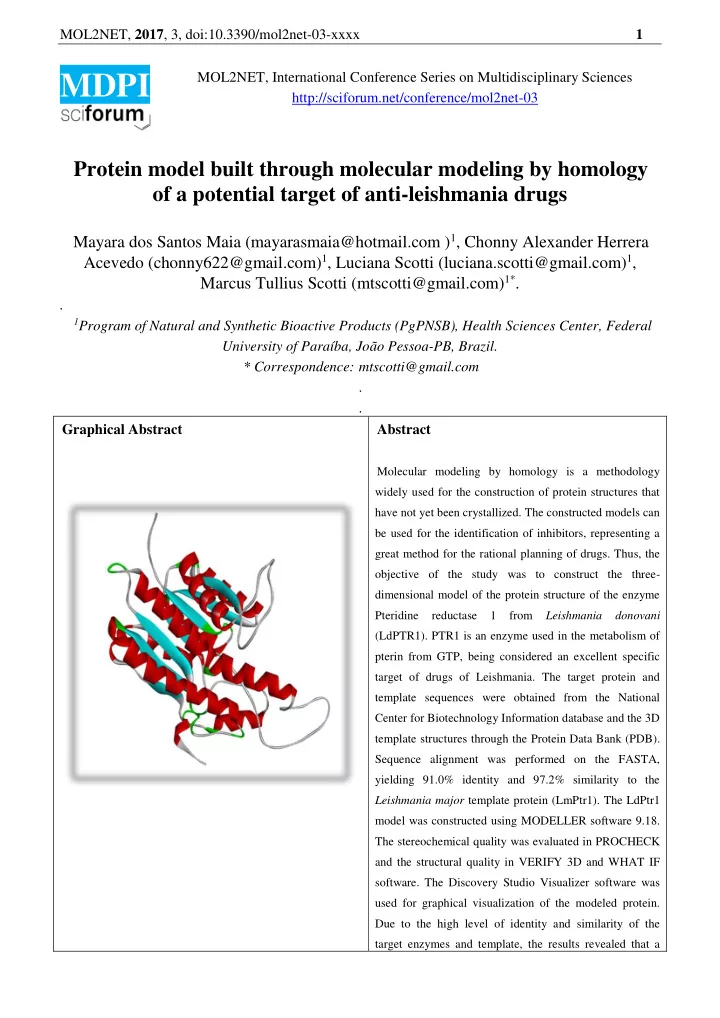
Molecular modeling by homology is a methodology widely used for the construction of protein structures that have not yet been crystallized. The constructed models can be used for the identification of inhibitors, representing a great method for the rational planning of drugs. Thus, the

  • bjective of the study was to construct the three-

dimensional model of the protein structure of the enzyme Pteridine reductase 1 from Leishmania donovani (LdPTR1). PTR1 is an enzyme used in the metabolism of pterin from GTP, being considered an excellent specific target of drugs of Leishmania. The target protein and template sequences were obtained from the National Center for Biotechnology Information database and the 3D template structures through the Protein Data Bank (PDB). Sequence alignment was performed on the FASTA, yielding 91.0% identity and 97.2% similarity to the Leishmania major template protein (LmPtr1). The LdPtr1 model was constructed using MODELLER software 9.18. The stereochemical quality was evaluated in PROCHECK and the structural quality in VERIFY 3D and WHAT IF

  • software. The Discovery Studio Visualizer software was

used for graphical visualization of the modeled protein. Due to the high level of identity and similarity of the target enzymes and template, the results revealed that a

slide-2
SLIDE 2

MOL2NET, 2017, 3, doi:10.3390/mol2net-03-xxxx 2

good model was constructed. The Ramachandran graph showed that the conformations of the main chain of amino acids are in allowed and favored regions. Besides that; 85.07% of the residues have the protein sequence compatible with their 3D structure and do not have unusual atomic contacts.

Introduction The development of a three-dimensional structural model of protein homologues can be used to identify inhibitors for specific targets by modeling homology and molecular dynamics. Homology modeling is currently the most accurate method, able to predict the structure of a protein based on the general observation that proteins with similar sequences possess. The computational methods for protein structure modeling play an important role in homology modeling [1]. Homology modeling requires the structure of an established protein (template) generated by a homologue containing the target sequence, provided that it shares approximately 30% or more similarity in the sequence or structure of the template [2]. Pteridine Reductase 1 (PTR1) is an enzyme used in the metabolism of pterin and necessary for the survival of the parasite [3]. Previous studies have reported as the PTR1 as an important target chemotherapeutic [3,4]. Inhibitors of Pteridine Reductase 1 (PTR1) proved to be lethal to the parasites in Leishmania

  • spp. and Trypanosoma brucei, but not in human

cells [4]. Thus, the objective of the study was to construct the three-dimensional model of the structure of the enzyme Pteridine reductase 1 from Leishmania donovani (LdPTR1) to contribute to the study of anti-leishmania inhibitors. Materials and Methods Identification of target sequences and selection

  • f protein template

The sequence of the target protein was

  • btained

from the National Center for Biotechnology Information database (https://www.ncbi.nlm.nih.gov/pubmed) and the identification of the resolved structures in 3D model candidates was done through a [5] and to

  • btain the structure, the RCSB Protein Data

Bank (https : //www.rcsb.org/pdb/home/home.do) [6]. The template protein selected was LmPtr1 (PDB ID: 1E7W). Alignment of template and target sequences Alignment of multiple sequences was performed using FASTA (http://www.ebi.ac.uk/Tools/sss/fasta/) and

  • btained the following values for Leishmania

donovani (LdPtr1): 91.0% identity and 97 , 2% similarity to Leishmania major Ptr1 (LmPtr1) (Figure 1). This was not the best score alignment in the FASTA, but it is the target organism of the study. Construction and validation of the model The LdPtr1 model was constructed using the homology molecular modeling method through MODELLER 9.18 software [7], which is based on the spatial constraints satisfaction

  • modeling. Five models were generated and the

lowest energy model was chosen. The stereochemical quality

  • fthe

model was evaluated in the PROCHECK [8], which

slide-3
SLIDE 3

MOL2NET, 2017, 3, doi:10.3390/mol2net-03-xxxx 3 evaluates several stereochemical parameters, such as torsional angles of the main chain, torsional angles of the side chain, bad contacts or steric impediments, flatness, among others. PROCHECK generates the Ramachandran graph [9] that verifies allowed and unallowed regions

  • f the main amino acid chain. The structural

quality was evaluated in VERIFY 3D software (http://services.mbi.ucla.edu/SAVES/) that analyzes the compatibility of the protein sequence with its 3D structure according to its chemical environments and WHAT IF (http: //swift.cmbi.ru.nl/servers/html/index.html) that analyzes various structural parameters, such as the atomic contacts between the residuals. The software Discovery Studio Visualizer [10] was used for graphic visualization of the modeled protein. Figure 1 - Alignment between the template sequence (LmPtr1) and the target sequence (LdPtr1). The regions highlighted in gray correspond to the identical amino acids in the two sequences, in yellow are the similar residues and in red the non-identical and non-similar residues. Results and Discussion Molecular Modeling by Homology of the enzyme Ptr1 of Leishmania donovani was performed using the Ptr1 enzyme of Leishmania major as template. A good LdPtr1 template was

  • btained because of the high level of similarity

between the target sequence (LdPtr1) and the template sequence (LmPtr1). To verify and validate the reliability and stereochemical quality

  • f

the modeled protein, data from the Ramachandran, VERIFY 3D and WHAT IF graph were considered. Analysis

  • f

the Ramachandran graph shows that the main chain conformations are 88.3% of the residues in the most favored regions, 11.7% allowed and 0%

  • utlier (Figure 2). In this analysis, since there

was no residue in the outlier region, the generated model was considered satisfactory. The G factors, which indicate the quality of covalent distance and bond angle, were 0.15 for dihedrons and 0.09 for phi / psi. Positive or slightly negative values indicate a model of good stereochemistry. Figure 2 - Ramachandran plot of LdPtr1 using Procheck. No VERIFY 3D; 85.07% of the residues had a mean 3D-1D score> = 0.2, which indicates

slide-4
SLIDE 4

MOL2NET, 2017, 3, doi:10.3390/mol2net-03-xxxx 4 a reliable model, since it is superior to 80% of the amino acids that marked = 0.2 in the 3D / 1D

  • profile. The quality of the atomic contacts

involving the atoms of each residue was analyzed using the Fine Packing Quality Control module

  • f the WHAT IF, which compares the

distribution of atom positions around each

  • residue. The mean score of all wastes is -0.687.

A score of less than -5.0 for a residue means bad

  • r unusual atomic contacts. The graphical

visualization of the modeled protein is observed in figure 3. Figure 3 - Graphical view of LdPtr1 in Discovery Studio Visualizer. Conclusions Molecular homology modeling is an excellent computational tool that performs the prediction

  • f

protein structures not yet crystallized, contributing to the rational planning

  • f anti-leishmania inhibitors. The LdPTR1 model

was considered satisfactory as observed in the validation results. References [1] KOPP J, SCHWEDE T. Automated protein structure homology modeling: a progress report. Pharmacogenomics 5(4):405–416, 2004. [2] CAVASOTTO, C. N; PHATAK, S. S. Homology modeling in drug discovery: current trends and applications. Drug Disc Today 14 (13–14): 676–683, 2009. [3] KHEIRANDISH, F.; BANDEHPOUR, M.; DAVOUDI N.; MOSAFFA, N.; DAWOOD, S.; KAZEMI, B.; HAGHIGHI, A.; KHAMESIPOUR, A.; MASJEDI, H.; MOHEBALI, M.; MAHBOUDI, F. Gene Regulation

  • f

Pteridine Reductase 1 in Leishmania Promastigotes and Amastigotes Using a Full-Length Antisense Construct. Iranian J Parasitol: Vol. 8, No.2, 2013. [4] PISA, F. D.; LANDI, G.; IACONO, L. D.; POZZI, C.; BORSARI, C.; FERRARI, S.; SANTUCCI, M.; SANTAREM, N.; CORDEIRO-DA-SILVA, A.; MORAES, C. B.; ALCANTARA, L. M.; FONTANA, V. FREITAS-JUNIOR, L. H.; GUL, S.; KUZIKOV, M.; BEHRENS, B.; PÖHNER, I.; WADE, R. C.; COSTI, M. P.; Mangani, S. Chroman-4-One Derivatives Targeting Pteridine Reductase 1 and Showing Anti-Parasitic Activity. Molecules, 22, 426, 2017. [5] ALTSCHUL, S. F.; GISH, W.; MILLER, W.; MYERS, E. W.; LIPMAN, D. J. Basic localalignment search tool, J. Mol. Biol. 215, 403–410, 1990. [6] BERNSTEIN, F. C.; KOETZLE, T. F.; WILLIAMS, G. J.; MEYER, E. F. J.; RICE B. R, M.D.; RODGERS, J. R.; KENNARD, O.; SHIMANOUCHI, T.; TASUMI, M. The Protein Data Bank: acomputer-based archival file for macromolecular structures, J. Mol. Biol. 112(3), 535–542, 1977. [7] SALI A, BLUNDELL T. Modelagem de proteína comparativa pela satisfação de restrições

  • espaciais. J Mol Bio. 234: 779-815, 1993.

[8] LAKOWSKI; R. A.; MACATHUR, M. W.; MOSS, D. S.; THORTON, J. M. PROCHECK: a program to check the stereochemicai quality of protein structures. J. Appl. Cryst. 26, 283-291, 1993. [9] LOVELL, S. C.; DAVIS, I. W.; ARENDALL III, W. B.; DE BAKKER, P. I. W.; WORD, J. M.; PRISANT, M. G.; RICHARDSON, J. S. AND RICHARDSON, D.

slide-5
SLIDE 5

MOL2NET, 2017, 3, doi:10.3390/mol2net-03-xxxx 5

  • C. Structure validation by Calpha geometry:

phi,psi and Cbeta deviation. Proteins: Structure, Function & Genetics. 50: 437-450, 2002. [10] DASSAULT SYSTÈMES BIOVIA, Discovery Studio Modeling Environment, Release 2017, San Diego: Dassault Systèmes, 2016.