SLIDE 1
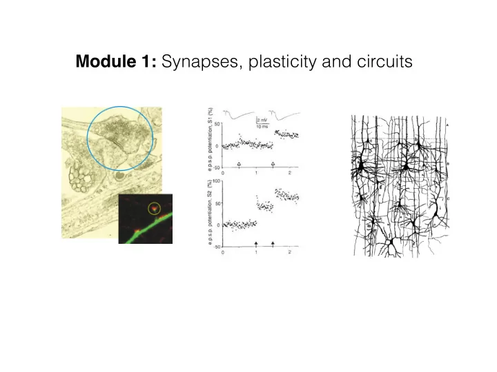
Module 1: Synapses, plasticity and circuits
SLIDE 2 The synapse: transfer of information
1 ms
SLIDE 3
The synapse: transfer of information
SLIDE 4
The synapse
SLIDE 5 Fatt and Katz, 1952
The miniature postsynaptic response (or ‘mini’)
- Remain in the presence of TTX
- Prolonged by blockers of acetylcholine esterase
- Blocked by AChR antagonists
SLIDE 6 Del Castillo and Katz, 1954
Quantal nature of neurotransmitter release
SLIDE 7
Quantal nature of neurotransmitter release
SLIDE 8 Quantal nature of neurotransmitter release
Heuser and Reese, 1981
Freeze fracture: vesicles caught in the act
SLIDE 9
Distinct vesicle pools Rapidly releasable pool Reserve Pool Resting Pool
SLIDE 10
The presynaptic vesicle cycle
SLIDE 11
SLIDE 12 Calcium Dependence of Neurotransmitter release
4- No calcium 1- No calcium 2- A little calcium 3- A little more calcium Katz
SLIDE 13
Caged-calcium experiments
SLIDE 14 Schneggenberger and Neher, nature 2000
Dependence of Neurotransmitter release on [Ca2+]int
SLIDE 15 Calcium nanodomains
Neher, CONB, 1998
SLIDE 16
SLIDE 17
Postsynaptic structures
SLIDE 18
Why spines?
1- Increase surface area to optimize packing of many synapses 2- Serve as a separate electrical unit that modulates synaptic signals 3- Provide a biochemical compartment that restricts mobility of molecules
SLIDE 19
Postsynaptic structure
Spines: occur at around 1-10 per um of dendrite
SLIDE 20 Synapse diversity: postsynaptic spine
Matsuzaki et al., 2001 Arellano et al., 2007
SLIDE 21 Postsynaptic structure: spines
Nimchinsky et al., ARN, 2002
SLIDE 22
Postsynaptic spine shape
Rneck = ⁄ "# $, where L is length of neck and A is cross-sectional area and " is resistivity of cytoplasm
SLIDE 23 Spine neck can filter synaptic events
Araya et al., PNAS, 2006
SLIDE 24 Noguchi et al., Neuron, 2005
Postsynaptic spine shape: calcium diffusion
SLIDE 25
Molecular architecture of excitatory synapses
SLIDE 26
SLIDE 27 Glutamate-gated channels
AMPAR NMDAR mGluRs
GluR1-4: Tetramers mostly of GluR2 and two others. Flip/flop: alternative splice variants Q/R editing: calcium permeability Almost all GluR2 subunits are in the R form, which is Ca2+ impermeable. GluN1-2: Tetramers of GluN1 (obligatory) and GluN2 A-D. Calcium permeable. Co-agonist: glycine. Blocked by Mg2+ at rest. 3 groups based on pharmacology Sequence and signalling. Group 1: mGlu1 and 5. Group 2: mGlu2 and 3. Group 3: mGlu4, 6, 7 and 8.
SLIDE 28
AMPA and NMDA currents
SLIDE 29
Na+ in K+ out
Normal situation recording Vm Inject current to depolarise to -20mV
Less Na+ in No ion movement
Inject more current
Na+ no mvt K+ out
Inject even more current ! No ion movement at the EPSP’s reversal potential
The EPSP: carried mainly by AMPA receptors
SLIDE 30
Glutamate postsynaptic currents
SLIDE 31
GABAA receptors
SLIDE 32
Cl- in
Normal situation recording Vm
No ion movement
Inject hyperpolarising current
Cl- Out
Inject more negative current
The IPSP
SLIDE 33
GABAB receptors
SLIDE 34
Plasticity of synapses and transmission: mechanisms
SLIDE 35
Short Term Plasticity: heterogeneous responses to spike trains
Same presynaptic neuron, different targets
SLIDE 36
Different presynaptic neurons, same target
Short Term Plasticity: heterogeneous responses to spike trains
SLIDE 37
Mechanisms: Possible Sites for Modulation
SLIDE 38 Width of an Action Potential
Geiger and Jonas, Neuron, 2000
SLIDE 39
Types of short-term plasticity
SLIDE 40
Facilitation at Granule to Purkinje Synapse
SLIDE 41
Facilitation and Residual Calcium
Could use slow buffer (eg: EGTA) to ‘mop up’ residual calcium
SLIDE 42 Facilitation and Residual Calcium
Alturi and Regehr, J. Neurosci., 1996
Process: high affinity, slow off rate
SLIDE 43
Plasticity of synapses and transmission: mechanisms and functional relevance
SLIDE 44 Carew and Kandel, 1973
SLIDE 45 Carew and Kandel, 1973
SLIDE 46
SLIDE 47 Bliss and Lomo, 1973
SLIDE 48 The Organisation of Behaviour (1949) When an axon of cell A is near enough to excite cell B and repeatedly or persistently takes part in firing it, some growth process or metabolic change takes place in one or both cells such that A's efficiency, as one of the cells firing B, is increased.[3] This is often paraphrased as "Neurons that fire together wire together." It is commonly referred to as Hebb's Law.
SLIDE 49 The Organisation of Behaviour (1949) When an axon of cell A is near enough to excite cell B and repeatedly or persistently takes part in firing it, some growth process or metabolic change takes place in one or both cells such that A's efficiency, as one of the cells firing B, is increased.[3] This is often paraphrased as "Neurons that fire together wire together." It is commonly referred to as Hebb's Law.
SLIDE 50
SLIDE 51
SLIDE 52
SLIDE 53