SLIDE 12 Acknowledgments
Prince Felipe Research Center Marie Curie Reintegration Grant STREP grant
COMPARATIVE MODELING Andrej Sali
Narayanan Eswar Min-Yi Shen Damien Devos Neboja Mirkovik Ursula Pieper MODEL ASSESSMENT Francisco Melo (CU) Alejandro Panjkovich (CU) FUNCTIONAL ANNOTATION Andrea Rossi Fred P. Davis MODELING ASSEMBLIES Frank Alber Fred P. Davis Maya Topf STRUCTURAL GENOMICS Stephen Burley (SGX) John Kuriyan (UCB) NY-SGXRC FUNCTIONAL ANNOTATION Fatima Al-Shahrour Joaquin Dopazo
Tropical Disease Initiative Stephen Maurer (UC Berkeley) Arti Rai (Duke U) Andrej Sali (UCSF) Thomas Kepler (Duke U) Ginger Taylor (TSL) EVA Burkhard Rost (Columbia U) Alfonso Valencia (CNIO)
BIOLOGY Jeff Friedman (RU) James Hudsped (RU) Partho Ghosh (UCSD) Alvaro Monteiro (Cornell U) Steven Krillis (St.George H)
CAMP Xavier Aviles (UAB) Hans-Peter Nester (SANOFI) Ernst Meinjohanns (ARPIDA) Boris Turk (IJS) Markus Gruetter (UE) Matthias Wilmanns (EMBL) Wolfram Bode (MPG)
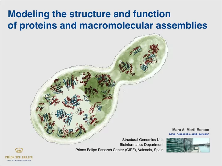
http://bioinfo.cipf.es/sgu/
University