
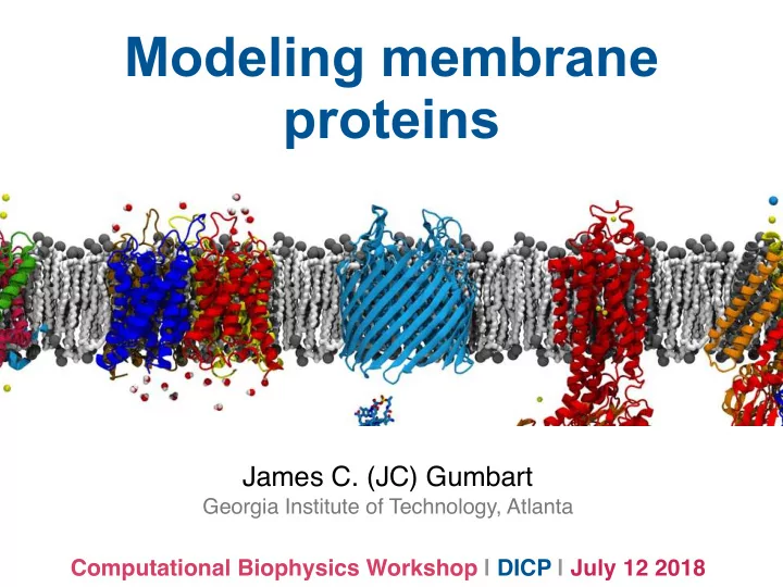
SLIDE 1
Modeling membrane proteins
James C. (JC) Gumbart
Georgia Institute of Technology, Atlanta
Computational Biophysics Workshop | DICP | July 12 2018
Modeling membrane proteins James C. (JC) Gumbart Georgia Institute - - PowerPoint PPT Presentation
Modeling membrane proteins James C. (JC) Gumbart Georgia Institute of Technology, Atlanta Computational Biophysics Workshop | DICP | July 12 2018 Why do living cells need membrane proteins? Living cells need to exchange materials and
Georgia Institute of Technology, Atlanta
Computational Biophysics Workshop | DICP | July 12 2018
Coarse-grained simulation of lipids randomly placed in water
~9 orders of magnitude slower ensuring bilayer asymmetry can be maintained
fluid mosaic model
Singer SJ, Nicolson GL (Feb 1972). Science 175: 720–31.
refined version (much more dense, varied)
β-barrel (outer membrane) α-helical (most membranes) many different ways to associate with membrane
receptors cell adhesion channels and transporters enzymes peripheral (not technically membrane proteins)
permit communication between outside and inside of the cell
nicotinic acetylcholine receptor
3) G-protein coupled, examples include rhodopsin, beta-2 adrenergic receptor (left) 2012 Nobel Prize in Chemistry (R.J. Lefkowitz, B.K. Kobilka) three classes: 1) enzyme linked, typically single TM 2) ligand-gated ion channels common example: neurotransmitter receptors (right)
G-protein GPCR
CAMs are on the cell surface, involved in binding to other cells intracellular domain interacts with the cytoskeleton extracellular domain interacts with other CAMs or EC Matrix conformational change initiated by signal from inside
communicate chemical, mechanical states transmembrane domain Example: integrin
Ex: cytochrome P450
substances
18,000 forms known
membrane-associated
reactions in drug metabolism
Cojocaru V, Balali-Mood K, Sansom MSP, Wade RC (2011) Structure and Dynamics of the Membrane-Bound Cytochrome P450 2C9. PLoS Comput Biol 7(8): e1002152.
typically only membrane anchored by a single TM examples include
and hydrolases
aquaporin, a water channel passive transport, solutes flow down (electro)chemical gradient most common solutes are ions
gramicidin, an unusual antibiotic ion channel KcsA, a bacterial K+ channel
Nobel Prize (2003) for channel structures, K+: R. MacKinnon; aquaporins: P. Agre
substrate binds from one side and releases to
couple the hydrolysis of ATP to drive transport structure of the Na+/K+ pump Examples include ion pumps, ATP synthase, ABC (ATP-binding cassette) transporters
Rahman KS, Cui G, Harvey SC, McCarty NA (2013) Modeling the Conformational Changes Underlying Channel Opening in CFTR. PLoS ONE 8(9): e74574.
Ex: homology model of Cystic Fibrosis Transmembrane Regulator (CFTR) evolved to be more channel-like (not strongly coupled) for Cl- Δ508 mutation found in 1/30 people, prevents expression in respiratory epithelial cells
ATP-binding domains transmembrane region
transport cycle for importer (exporter slightly different)
In the plasma membrane of animal cells, Na+ is the usual co-transported ion in bacteria/yeast (and organelles!) often H+
Science, 301:610-615, 2003.
(SGLT) in the kidneys
substrate → symporter
antiporter
transport energy comes from co-transport of an ion
transporter cycles through a number
three primary states: 1) outward open 2) occluded 3) inward open no transporter has structures in ALL states
■ Molecular dynamics simulations of membrane channels and transporters.F Khalili-Araghi, J Gumbart, P-C Wen, M Sotomayor, E Tajkhorshid, and K Schulten. COSB, 19:128-137, 2009.
glycolipid transfer protein phospholipase A2 - involved in lipid metabolism, also in many venoms (promotes cell lysis) enzymes hydrophobic molecule transporters GLA domain - involved in blood coagulation cascade
Tajkhorshid Lab (UIUC): N. Tavoosi,et
structural
Tajkhorshid Lab (UIUC): N. Tavoosi,et al. (2011) JBC. 286: 23247.
potential of mean force (PMF) projects full free-energy space onto one (or more) selected reaction coordinates
W(z) = −kT ln(P(z) P0 )
also can be expressed in terms probability: 2D PMF for ion transport through gramicidin A knowledge of PMF permits determination of many properties, e.g., conductance, average times, binding sites, etc.
Conduction in the Gramicidin A Channel.Proc. Nat. Acad. Sci. 101, 117-122.
GypA-E expressed at surface of red blood cells, acts as a receptor, prevents aggregation, etc. NMR structure of TM domain only PMF for helix-helix association in membrane as function of separation distance dimer is favored by 10 kcal/mol
by GxxG motif
Henin, J.; Pohorille, A.; Chipot, C. Insights into the recognition and association of transmembrane alpha-helices. The free energy of alpha-helix dimerization in glycophorin A JACS 2005, 127 (23), 8478-8484.
AmtB - an ammonia (NH3)/ammonium (NH4+) channel homologous to RhxG (x=A,B,C) proteins in mammalian blood cells channel is hydrophobic - NH4+ likely changes protonation states at entrance/ exit PMF for NH3 moving through channel shows minima at crystallographically resolved binding sites determined using adaptive biasing forces (ABF) in NAMD
Step 1: Get the protein PDB from the PDB databank Step 2: Build a PSF, including repeated subunits if necessary Step 3: Build the membrane, using VMD (POPE, POPC only) or CHARMM-GUI Step 4: Orient the protein in the membrane and combine them, removing overlapping lipids - write a new PSF/PDB Step 5: Add water above and below using VMD Solvate, removing any that accidentally get placed inside the membrane Step 6: Add ions; prepare inputs for minimization and equilibration
Go to the Orientations of Proteins in Membranes (OPM) database Look up your protein to see the details of its multimeric state,
http://opm.phar.umich.edu/
CHARMM-GUI can read the aligned, multimeric protein directly from OPM and build the membrane, water, and ions around it
http://charmm-gui.org/
Ex: AmtB (PDB 1U7G) an NH3/NH4+ channel OPM shows that it is a trimer CHARMM- GUI can take output from OPM directly
Think carefully about what to include! Three copies of the protein Crystallographic water NH3/NH4+ (substrates
BOG: β-octylglucoside (detergent for crystallization)
There are a number of other choices to make along the way For example, how to patch the termini of the proteins NTER and CTER usually appropriate
Which lipids to use for the membrane? Ideally, want to select lipids to match the native membrane composition! Search textbooks, papers, etc. for estimates of the lipids and ratios for the membrane of interest
Gram-negative outer membrane mammalian plasma membrane mitochondrial membrane Gram-negative inner membrane
For simplicity, used single- component POPE here Which lipids to use for the membrane?
The system looks reasonable overall, but there are a few potential problems After a few more choices, a complete system is output (step 5)
Initial guess of membrane size (100 Å x 100 Å) is too small want to have more than 2-3 layers
images
shrink after equilibration! (need 30 Å at least after eq.)
These are not the fault of CHARMM-GUI! Instead they are co-crystallized waters Option 1: rebuild but leave these boxes un-checked Option 2: use a script to delete intra- membrane waters (see tutorial)
If you look closely at the waters, they look very strange! Why is there a third bond???
When adding water with VMD Solvate, that extra bond is commented
NAMD doesn’t care about it (it is harmless, just ugly!)
Warning: Ignored 19521 bonds with zero force constants. Warning: Will get H-H distance in rigid H2O from H-O-H angle.
First, relax lipid tails for (water/prot/lipid heads restrained) for ~0.5 ns Second, relax lipids and water (protein restrained) for 3-5 ns to ensure a good packing of lipids around the protein Finally, can run with everything released in NpT ensemble NOTE: CHARMM27 lipids do not maintain correct area/lipid but CHARMM36 lipids do! Always use the latest force field! System has to be relaxed carefully to avoid distortions