
SLIDE 1
[ADD PRESENTATION TITLE: INSERT TAB > HEADER & FOOTER > NOTES AND HANDOUTS] 12/11/2015 1
A rational approach to knee pain Primary Care Sports Medicine 2015
12/11/2015
Brian Feeley, MD Associate Professor, Sports Medicine and Shoulder Surgery UCSF Department of Orthopaedic Surgery
Top 5 4 knee problems:
12/11/2015
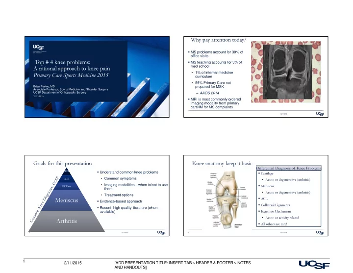
Why pay attention today?
MS problems account for 30% of
- ffice visits
MS teaching accounts for 3% of med school
- 1% of internal medicine
curriculum
- 56% Primary Care not
prepared for MSK ‒ AAOS 2014 MRI is most commonly ordered imaging modality from primary care/IM for MS complaints
Goals for this presentation
Understand common knee problems
- Common symptoms
- Imaging modalities—when to/not to use
them
- Treatment options
Evidence-based approach Recent high quality literature (when available)
12/11/2015 3
MCL MCL ACL ACL PF Pain PF Pain
Meniscus Meniscus Arthritis Arthritis
Knee anatomy-keep it basic
12/11/2015 4
Cartilage
- Acute or degenerative (arthritis)
Meniscus
- Acute or degenerative (arthritis)
ACL Collateral Ligaments Extensor Mechanism
- Acute or activity related
All others are rare!