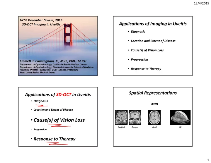
Cause(s) of Vision Loss Sagittal Coronal Axial 3D Progression - - PowerPoint PPT Presentation

cause s of vision loss
SMART_READER_LITE
LIVE PREVIEW

Cause(s) of Vision Loss Sagittal Coronal Axial 3D Progression - - PowerPoint PPT Presentation

12/4/2015 UCSF December Course, 2015 Applications of Imaging in Uveitis SD-OCT Imaging in Uveitis Diagnosis Location and Extent of Disease Cause(s) of Vision Loss Progression Emmett T. Cunningham, Jr., M.D., PhD., M.P.H


slide-1
SLIDE 1

12/4/2015 1 Emmett T. Cunningham, Jr., M.D., PhD., M.P.H

Department of Ophthalmology, California Pacific Medical Center Department of Ophthalmology, Stanford University School of Medicine Francis I. Proctor Foundation, UCSF School of Medicine West Coast Retina Medical Group

UCSF December Course, 2015 SD-OCT Imaging in Uveitis

Applications of Imaging in Uveitis

  • Diagnosis
  • Location and Extent of Disease
  • Cause(s) of Vision Loss
  • Progression
  • Response to Therapy

Applications of SD-OCT in Uveitis

  • Diagnosis
  • Location and Extent of Disease
  • Cause(s) of Vision Loss
  • Progression
  • Response to Therapy

Spatial Representations

Axial Coronal Sagittal 3D

MRI

slide-2
SLIDE 2

12/4/2015 2 Line Scans En Face 3D

Horizontal Vertical

Spatial Representations

Horizontal Vertical En Face

(Retinal Thickness; ILM – RPE)

SD-OCT Representations

3D

Slab ILM-based Slab RPE-based Slab

Anatomical En Face Imaging

Courtesy Dr. Brandon Lujan

8 Fovea (“pit”)

SD-OCT = Virtual Histology < 10 µm resolution

Courtesy of Ralph Eagle, MD Wills Eye Hospital

slide-3
SLIDE 3

12/4/2015 3

International Nomenclature for OCT Meeting Consensus Normal OCT Terminology

Formed Vitreous Posterior Cortical Vitreous

Preretinal Space Nerve Fiber Layer Ganglion Cell Layer Inner Plexiform Layer Inner Nuclear Layer Outer Plexiform Layer (dendritic) Henle’s-ONL junction (subtle) Henle Fiber Layer (axonal OPL) Outer Nuclear Layer Sattler’s Layer (Inner Choroid) Haller’s Layer (Outer Choroid) External Limiting Membrane Ellipsoid Zone (EZ) Outer Segments Interdigitation Zone (IZ) Choriocapillaris RPE/ Bruch’s Complex Myoid Zone Choroid Sclera Junction

Normal SD-OCT ‘The Essentials’

Sclera = 1000µm Choroid Normal Lacunae – No Infiltration or Masses 250 µm 250 µm

Normal SD-OCT ‘The Essentials’

Sclera = 1000µm Choroid Normal Lacunae – No Infiltration or Masses

GCL INL ONL

NFL IPL OPL

3 Nerve Fiber Layers 3 Nuclear Layers 250 µm 250 µm

Normal SD-OCT ‘The Essentials’

Sclera = 1000µm Choroid Normal Lacunae – No Infiltration or Masses

GCL INL ONL

NFL IPL OPL

3 Nerve Fiber Layers 3 Nuclear Layers 4 Outer Retinal Bands

PR-RPE Complex

250 µm 250 µm

slide-4
SLIDE 4

12/4/2015 4

Diagnosis

Plus those not listed . . .

Dozens of Disorders

Syphilis Syphilis

20/80

slide-5
SLIDE 5

12/4/2015 5

SRF

Vitreous Cells Nodular RPE Nodular RPE

20/80 20/80 20/80 En Face PR Layer 20/80

slide-6
SLIDE 6

12/4/2015 6 20/80 FAF 20/80

1:10.87

20/80

5:46.90

20/80 Acute Syphilitic Posterior Placoid Chorioretinitis

slide-7
SLIDE 7

12/4/2015 7

Location and Extent of Disease

APMPPE APMPPE

OD 20/16 OD 20/16

Disruption / Hyper-reflectivity Outer Retina

slide-8
SLIDE 8

12/4/2015 8 OD 20/16 En Face PR Layer

Placoid Lesions

OS 20/16 OD 20/16 Acute Mulifocal Placoid Pigment Epitheliopathy

http://westcoastretina.com/august-2013.html

Case of the Month

Acute Mulifocal Placoid Pigment Epitheliopathy AMPPE

Early Hypo-FA, Late Hyper-FA

slide-9
SLIDE 9

12/4/2015 9

Cause(s) of Vision Loss

Sarcoidosis Sarcoidosis

OS 20/25-1 OS 20/25-1

slide-10
SLIDE 10

12/4/2015 10

OS 20/25-1 OS 20/25-1

INL Cysts HFL Cysts

En Face Henle Fiber Layer OS 20/25-1 En Face INL OS 20/25-1

slide-11
SLIDE 11

12/4/2015 11

Progression

BSCR BSCR

20/30+2 HLA-A29+ BSCR 20/30+2

00:20.29

BSCR “Dry type”

slide-12
SLIDE 12

12/4/2015 12

20/30+2

07:52.73

BSCR “Dry type” 20/30+2 At Presentation 20/50-2 + Two Years Lost to Follow Up No Treatment 20/50-2 + Two Years Lost to Follow Up No Treatment 20/30+2 Baseline

slide-13
SLIDE 13

12/4/2015 13

Response to Therapy

VKH VKH

SRD SRD

PR-RPE Adhension Convex RPE Vitreous Cells

20/125

slide-14
SLIDE 14

12/4/2015 14

2:23.93

20/125

7:06.84

20/125 Vogt-Koyanagi-Harada Disease 20/125 20/25

Three Months After Initiating High-Dose Prednisone

slide-15
SLIDE 15

12/4/2015 15

Hashizume

Hashizume et al, Long-term Observation Of Choroidal Thickness In Vogt-Koyanagi-Harada Disease. ARVO 2012; Program / Poster 3203/A48.

Applications of SD-OCT in Uveitis

  • Diagnosis
  • Location and Extent of Disease
  • Cause(s) of Vision Loss
  • Progression
  • Response to Therapy

emmett_cunningham@yahoo.com