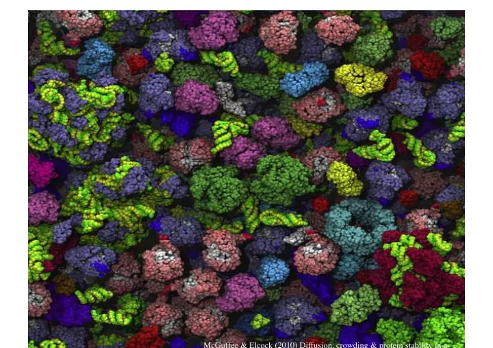
SLIDE 1 McGuffee & Elcock (2010) Diffusion, crowding & protein stability in a
SLIDE 2
Macromolecular Crowding
SLIDE 3 Molecules and Complexes: E. coli Census
- Nproteins ~ 3x10^6
- Nribosomes ~ 20,000
- Nlipids ~ 2x10^7
Spacing for a molecule of given concentration c:
d = c−1 3
SLIDE 4 Protein Spacing in E. coli
- Estimate
- Average protein-protein distance ~ 10 nm
- 1 mM protein in vitro distance ~ 100 nm
Radius of protein ~ 2nm 1 um
SLIDE 5 Linked Polymer Network Architecture
Gram positive bacterial wall (atomic resolution) Collagen fibrils Basement membrane Actin web in a miving cell Cellulose network
Ligament collagen bundle Axon bundle Intenstinal microvilli
SLIDE 6
- Effect on binding and interaction
- Difference between cells and dilute
solutions
SLIDE 7 Ten Commandments of Enzymology, Amended
Kornberg (2003) Tr. Biochem. Sci.
- 1. Thou shalt rely on enzymology to resolve
and reconstitute biologic events
- 2. Trust the universality of biochemistry and
the power of microbiology (Escherichia sapiens)
- 3. Not believe something just because you can
explain it
- 4. Not waste clean thinking on dirty enzymes
- 5. Not waste clean enzymes on dirty substrates
- 6. Use genetics and genomics
- 7. Be aware that cells are molecularly crowded
- 8. Depend on viruses to open windows
- 9. Remain mindful of the power of radioactive
tracers
- 10. Employ enzymes as unique reagents
Arthur Kornberg Nobel Prize for Chemistry (1959) Discovery of DNA polymerase (now known as DNA polymerase I)
Be aware that cells are molecularly crowded
SLIDE 8 Fish skin keratocyte
Fish Keratocyte Leading Edge
Membrane stripped platinum coated E.M. Front edge: Ordered, Branched Middle zone: Randomly
filaments
SLIDE 9 Cellular Effects of Crowding
- Equilibrium binding
- Diffusive processes
SLIDE 10 7.5% 5% 2.5% 0% PEG concentration
ATPase Rate
T4 DNA polymerase clamp-loader proteins
SLIDE 11 Experimental Measures
BODIPY-FI sizes
Verkman (2002) TiBS
Method 1 Method 2 FCS Time resolved Fluorescence Anisotropy Method 3
SLIDE 12 Cellular Diffusion Coefficients
Verkman (2002) TiBS
SLIDE 13 Single Molecule Imaging Membrane Proteins
GFP-Lck Lck Tyrosine kinase Jurkat T-cells TIRF microscopy Anti-T cell receptor Abs stimulate clustering
Douglass & Vale (2005)
Trapped Long distance
SLIDE 14 Membrane Microdomains
T-cell Signalling domains in CD2-enriched signalling domains Signalling activated by antibody-patch on coverslip
CD2 Lck Merge
SLIDE 15 Lipid Rafts
GPI-anchored proteins Raft-associated lipids
Mayor Lab, NCBS Bangalore
SLIDE 16 Effective Diffusion
<d^2> = < [d(t)-d(t+δt)]^2 >, For different values of δt. Plotting d^2 vs δt gives us a profile that can be fitted by <d^2>=2*D*t^α α = 1 for normal diffusion
Bacher, Reichenzeller, Athale et al. (2004) Klopfstein et al. (2002) Cell
Raft targeted Lck-10 GFP Lck GFP
SLIDE 17 TIRF: At the Surface
Evanescent wave illumination with limited range
Nikon Instruments
SLIDE 18 Lattice Model of Crowding
Ωv = reaction volume L=ligand no. C=Crowding
SLIDE 19 Ligand-Receptor Binding pbound = 1 1+ Ω− L − C L
( )eβΔε L
L << Ω When C increases, pbound increases
ΔεL = εL
b −εL sol
SLIDE 20
Crowding Changes L-R binding Probability
SLIDE 21 7.5% 5% 2.5% 0% PEG concentration Binding constant PEG dependent)
Dissociation Rate
Kd = 1 v eβΔe
pbound = L
[ ] Kd
( )
n
1+ L
[ ] Kd
( )
n
v=volume of single lattice site Δε = binding energy
SLIDE 22
PEG as Crowdant
T4 atpase data, PEG size (12 kDa) << Protein size (164 kDa) Ω large boxes r small boxes in each large box
SLIDE 23
Crowdant Smaller than Ligand
SLIDE 24
Binding
Where, volume fraction of the crowding molecules in solution Assuming L << Ω And (N+r)!/N! ~ Nr
φC = C rΩ
pbound = 1 1+ Ω L (1− φC )reBΔε L
SLIDE 25
Dissociation Constant
Volume fraction dependent dissociation constant Kd
Kd φC
( )
Kd φC = 0
( )
= 1− φC
( )
r
Kd = 1 v eβΔe
SLIDE 26
Factors Affecting Crowding
SLIDE 27
Osmotic Pressure and Crowding
Osmotic pressure due to excess Hemoglobin p = pressure v=volume of single box in lattice [H]=concentration of Hb molecules=H/Ωv
p = − kBT v ln(1−[H]v)
SLIDE 28 Crowding and Osmotic Pressure
Free parameter is v V=5.8 nm Hard sphere gas model Experiment Lattice gas
SLIDE 29
Hard Sphere Gas Model
Boltzmann 1899 Where x = 4V[H] V=volume of hard sphere
p = kBT[H](1+ x + 0.625x 2 + 0.287x 3 + 0.11x 4)
SLIDE 30 Next
- Crowded polymers and ordering
- Cytoskeleton
- Motors
- Cells in tissues
- FRAP data
- Paper presentations
SLIDE 31
2011-03-23
SLIDE 32 Macromolecular Crowding
- 10-100% of fluid volume of cytoplasm lies
within 1 molecular diameter of the surface
- f fibrous and membraneous structures
- Pores
- Reactant X and pore size comparable
SLIDE 33 Sieving Effect
Verkman (2002) TiBS
SLIDE 34 Volume Exclusion
Minton (2001) J. Biol. Chem.
Excluded volume Available volume
SLIDE 35 Minton (2001) J. Biol. Chem.
Relative Sizes
SLIDE 36
Volume Available
Effective and actual concentrations vtot=total volume va,i=volume available to species i
γ i ≡ ai ci
( ) = vtot va,i
( )
SLIDE 37 Haemoglobin Concentration
Normal RBC Concentration ~ 300gm/L
SLIDE 38
2011-03-29
SLIDE 39 Forces due to Volume Exclusion
Large particle near surface Two large particles in solution Two rod-like molecules in solution Depletion forces Volume available to smaller molecules increases
SLIDE 40
Origin of Depletion Forces
R= radius of large disk r = radius of small disk Surface 2D geometry Find area available to small disks, as a function of distance z between large disk and surface
SLIDE 41 Excluded Volume Interactions
R= radius of large disk r = radius of small disk Surface 2D geometry z=distance between large disk and surface
Area available to small disks Entropy increases
SLIDE 42 Free Energy Change
No conventional forces- van der Walls, electrostatics, etc. Free energy change by change in entropy is: Vbox = volume of box, Vex=excluded volume, v=volume
- f unit cell, N=no. of SMALL MOLECULE particles
Gex = −NkBT ln Vbox −Vex v + NkBT ln Vbox v
SLIDE 43
Free Energy
If Vex << Vbox, approximate ln(1+x) ≈ ln(x) If 2 large particles overlap excluded volumes, Vex increases entropy of small particle ~ ideal gas (osmotic) pressure of small particles in box
Gex = NkBT Vex Vbox
NkBT Vbpx
SLIDE 44
Depeletion Force
SLIDE 45 Volume and Force
Total excluded volume Volume of spherical cone Volume cone Overlap Depletion Force
Vex = 2⋅ 4π 3 R + r
( )
3 −Voverlap
Vsphericalcone = 2π 3 R + r
( )
2 ⋅ R + r − D 2
( )
Vcone = π 3 D 2 R + r
( )
2 − D 2
( )
2
[ ]
Voverlap = 2π 3 R + r + D 2
( )
2 2R + 2r + D 2
( )
Fdepletion = −∂Gex ∂D = −pπ R + r
( )
2 − D2
4
p = nkBT , n=N/Vbox, and distance 2R<D<2(R+r)
SLIDE 46 Depletion Force Measurement
2 beads, R=625 nm, Move in line Depletion agent- Phage λ DNA r=500 nm
DNA conc. DNA conc. DNA conc.
p(D) ∝e−βGex (D)
Gex(D) = pVoverlap
SLIDE 47 Entropic Ordering
Rods: Filamentous viruses Spheres
SLIDE 48
Volume Exclusion
Mutual exclusion v=volume occupied N=number of macromolecules Ω=total number of boxes N=no. of macromolecules In absence of excluded volume Zex(N) = Ω! N! Ω− N
( )!
Znex(N) = ΩN N!
SLIDE 49 Free Energy for Excluded Volume
Free energy Using stirling’s approximation, and assuming Ω >> N, and
G = −kBT lnZ
ΔGex = Gex − Gnex = −kBT ln Zex Znex
1− N Ω
( )
Ω ≈ e−N
Zex Znex ≈ 1− N Ω
N
ΔGex = −NkBT ln 1− N Ω ≈ kBT N 2 Ω
SLIDE 50 Polymers, Crowding and RW Model
- Random walk model ignores self-avoidance
- Size of macro-molecule like DNA ~
N=number of segments, a=persistence length
Na2
Competing effects:
- Entropy makes chain compact
- Self-avoidance swells the chain
SLIDE 51
Random Walk Polymer
Free energy R=radius of polymer SRW(R)=random walk entropy of chain of length R Entropy from probability distribution P(R;N)
G(R) = −TSRW (R) + Gex(R)
SRW (R) = kBT lnP(R;N) + const = −kB 3R2 2Na2 + const
SLIDE 52 Excluded volume
- Assume polymer to be gas with hard cylinders
- f length a and diameter d
- Mutual orientation angle θ decides excluded
volume v = 2da2 sinθ
SLIDE 53
- For d<<a
- Averaging sin(θ) over all orientations gives
estimate for excluded volume
- Free energy
- Volume fraction of N hard cylinders
πa2d 2
Gex = kBTNφ
φ(R) = N 1 2 πa2d 1 2 πR3 = N 3a2d 8R3
SLIDE 54 Free Energy Difference
- Since
- Flory’s estimate of free energy of polymer
- Size of chain
ΔGex ≈ kBT N 2 Ω = kBTNφ Gex(R) = kBTN 2 3a2d 8R3 G flory(R) = kBT 3R2 2Na2 + kBTN 2 3a2d 8R3
Rflory = 3 8 a4d
1/ 5
N 3/ 5
SLIDE 55 RW vs. SAW
- Power scaling RW (N1/2), SAW (N3/5)
- Short polymers RW still valid
For DNA d~2nm, a=100nm For N<<16(d/a)=40,000, G self avoid < RW entropy L=Na=40,000x100 nm (kuhn length) ~ 16 µm
Gex = kBT 3 8 d a N
1 2
SRW = 3 2 kBT
SLIDE 56
Protein Folding & Crowding
Chaperones
SLIDE 57
Diffusion in Crowded Environs
SLIDE 58 Diffusion in Crowded Environments
- For single time step t=τ
- After time t, steps N=t/τ, so
- Diffusion coefficient
For a random walker
x 2 τ = a2 ⋅ pright + a2 ⋅ pleft + 0⋅ pstay = a2(1− φ) x 2 t = t τ x 2 τ = a2 τ (1− φ)t
D = D0(1− φ)
D0 = a2 2τ
SLIDE 59 FITC-Aldolase diffusing
Aldolase BSA Ovalbumin
SLIDE 60
Self-Diffusion and Crowding
SLIDE 61 NEXT
- Cytoskeleton and motors
- Cells
- Development
SLIDE 62
SLIDE 63