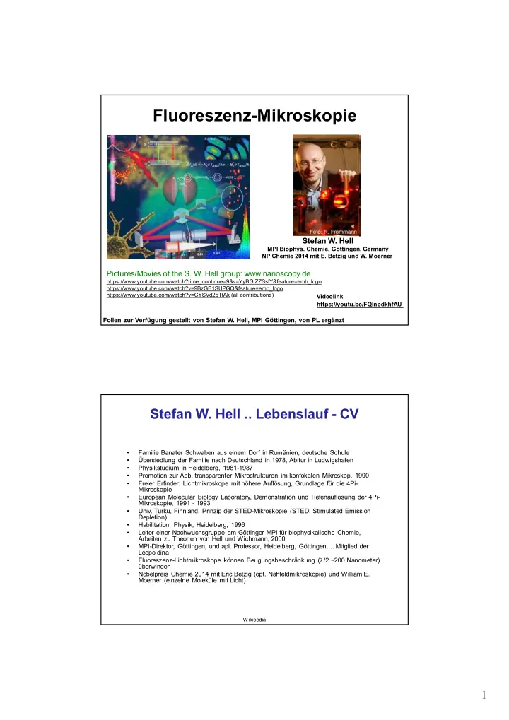
Fluoreszenz-Mikroskopie Stefan W. Hell MPI Biophys. Chemie, - - PDF document

fluoreszenz mikroskopie
SMART_READER_LITE
LIVE PREVIEW

Fluoreszenz-Mikroskopie Stefan W. Hell MPI Biophys. Chemie, - - PDF document

Fluoreszenz-Mikroskopie Stefan W. Hell MPI Biophys. Chemie, Gttingen, Germany NP Chemie 2014 mit E. Betzig und W. Moerner Pictures/Movies of the S. W. Hell group: www.nanoscopy.de


slide-1
SLIDE 1

1

Stefan W. Hell

MPI Biophys. Chemie, Göttingen, Germany NP Chemie 2014 mit E. Betzig und W. Moerner

Fluoreszenz-Mikroskopie

Folien zur Verfügung gestellt von Stefan W. Hell, MPI Göttingen, von PL ergänzt

Pictures/Movies of the S. W. Hell group: www.nanoscopy.de

https://www.youtube.com/watch?time_continue=9&v=YyBGiZZSslY&feature=emb_logo https://www.youtube.com/watch?v=9BzGB1SUPGQ&feature=emb_logo https://www.youtube.com/watch?v=CYSVd2qTfAk (all contributions) Videolink https://youtu.be/FQlnpdkhfAU

Stefan W. Hell .. Lebenslauf - CV

  • Familie Banater Schwaben aus einem Dorf in Rumänien, deutsche Schule
  • Übersiedlung der Familie nach Deutschland in 1978, Abitur in Ludwigshafen
  • Physikstudium in Heidelberg, 1981-1987
  • Promotion zur Abb. transparenter Mikrostrukturen im konfokalen Mikroskop, 1990
  • Freier Erfinder: Lichtmikroskope mit höhere Auflösung, Grundlage für die 4Pi-

Mikroskopie

  • European Molecular Biology Laboratory, Demonstration und Tiefenauflösung der 4Pi-

Mikroskopie, 1991 - 1993

  • Univ. Turku, Finnland, Prinzip der STED-Mikroskopie (STED: Stimulated Emission

Depletion)

  • Habilitation, Physik, Heidelberg, 1996
  • Leiter einer Nachwuchsgruppe am Göttinger MPI für biophysikalische Chemie,

Arbeiten zu Theorien von Hell und Wichmann, 2000

  • MPI-Direktor, Göttingen, und apl. Professor, Heidelberg, Göttingen, .. Mitglied der

Leopoldina

  • Fluoreszenz-Lichtmikroskope können Beugungsbeschränkung (l/2 ~200 Nanometer)

überwinden

  • Nobelpreis Chemie 2014 mit Eric Betzig (opt. Nahfeldmikroskopie) und William E.

Moerner (einzelne Moleküle mit Licht)

Wikipedia

slide-2
SLIDE 2

2

Stefan W. Hell, MPI Göttingen

200 nm

Lens

500 nm

Wavelength

 l   sin n 2 x

l 

  • E. Abbe (1873), Arch. Mikroskop. Anat. 9, 413.

Die Qual mit der Auflösung:

  • optische Mikroskopie ist praktisch
  • es gibt lange Erfahrung mit optischen Instrumenten
  • weit verbreitet: das Auge, Experimente sind möglich an Luft, ..
  • Absorption organischer Materie ist klein im sichtbaren Spektralbereich

Stefan W. Hell, MPI Göttingen

Verschiedene Lösungsmöglichkeiten: Beugung ist eine fundamentale Eigenschaft der EM-Welle

  • konfokale Mikroskopie

– Beleuchtung und Bildgebung geschehen durch beugungsbegrenzte Foki – Rastermethode: Optimierung und Ortsinformation

  • Immersionsmikroskopie

– Flüssigkeit (n>1) zwischen Objekt und Objektiv

  • 4pi-Mikroskopie

– Vergrößerung des Raumwinkels

  • Fluoreszenzspektrokopie

– Farbstoffen werden an selektiven Plätzen gebunden/eingebaut – Farbstoffen werden über Laser gezielt ein/aus geschaltet – Zeit- und Frequenzauflösung: Dynamik und Struktur (Ort) Nähe des Farbstoffs

  • FRED: Resonanter Fluoreszenz Energie Transfer nach Förster

– Energietransfer zwischen Sender- und Empfängermolekül hängt vom Abstand ab – bestimme die Zerfallszeit des Senders -> Abstand – Zeit- und Ortsinformation

  • Nahfeldoptik

– benutzt evaneszente Wellen an der Oberfläche, z.B. durch Totalreflexion – örtliche Einschränkung

slide-3
SLIDE 3

3

Stefan W. Hell, MPI Göttingen

4Pi- Microscopy: resolution improvement in Z

Stefan W. Hell, MPI Göttingen

4Pi- Microscopy:

70 - 140 nm

     

4 1 2

, , , , , ,

Pi

E r z E r z E r z       r r r

Coherent illumination and/or fluorescence detection

S.W. Hell (1990), Europ. Patent OS 0491289. S.W. Hell, et al. (1992), Opt. Commun. 93, 277.

  • M. Schrader, et al. (1998), Biophys. J. 75, 1659.
  • H. Gugel, et al. (2004), Biophys. J. 87, 4146.
slide-4
SLIDE 4

4

Stefan W. Hell, MPI Göttingen

2 µm 2 µm

Confocal 4Pi

Z X

Microtubules, mouse fibroblast Immunofluor, Oregon Green

S.W. Hell, et al. (1992), Opt. Commun. 93, 277.

  • M. Schrader, et al. (1998), Biophys. J. 75, 1659.
  • H. Gugel, et al. (2004), Biophys. J. 87, 4146.

Stefan W. Hell, MPI Göttingen

Commercial 4pi-microscope

Z- resolution < 90 nm (Live cells /aqueous cond.)

  • H. Gugel, et al. (2004), Biophys J 87, 4146.
slide-5
SLIDE 5

5

Stefan W. Hell, MPI Göttingen

STED-Mikroskope Stimulierte Emission und Ausbleichung

1st physical concept to break the diffraction barrier in far-field fluorescence microscopy

S.W. Hell & J. Wichmann (1994), Opt. Lett. 19, 780.

  • Fluoreszenzfarbstoffe als Marker
  • Anregung durch Licht bestimmter Wellenlängen
  • spontane Abstrahlung: Fluoreszenz
  • stimulierte Emission schalten die Moleküle wieder aus

Stefan W. Hell, MPI Göttingen

Dynamik von Farbstoffmolekülen

  • angeregtes Farbstoffmolekül kann durch FL oder

stimulierte Emission in den Grundzustand übergehen

  • Bestrahlung des angeregten Farbstoffmolekül

vergleichbarer Wellenlänge wie Fluoreszenzlichts

  • Übergang in den Grundzustand durch Aussenden

eines Photons

  • spontane Fluoreszenz ist danach unterdrückt
  • Prozesse werden durch Farbfilter getrennt

S0 S

1

Absorption Stimulated Emission

Fluorescence 1 ps

vib

 p 1 s

fl

n  

200 nm x y S.W. Hell & J. Wichmann (1994), Opt. Lett. 19, 780.

slide-6
SLIDE 6

6

Stefan W. Hell, MPI Göttingen

Dynamik von Farbstoffmolekülen

STED-Mikroskop:

  • Verbesserung der konfokalen Mikroskopie (Rastern)
  • Reduktion des Fluoreszenzbereichs unter den

Bereich der Beleuchtung durch gezieltes Ausschalten im Außenbereich des Fokus

  • zweiter Strahl hat ringförmiges Profil und ist dunkel

wo der Anregungsstrahl maximal ist

  • wenige Photonen reichen zur Stimulation
  • der nicht ausgeschaltete zentrale Bereich ist sehr viel

kleiner

  • Scannen der Probe

Stefan W. Hell, MPI Göttingen

STED-Mikroskopie

3 6 9 1.0 0.5 0.0

ISTED [GW/cm2]

Fluorescence

Detector

y x z

Excitation Depletion (STED)

Phase Modulation

50 ps 50-200 ps S.W. Hell & J. Wichmann (1994), Opt. Lett. 19, 780. The stronger the STED beam the narrower the fluorescent spot!

slide-7
SLIDE 7

7

Stefan W. Hell, MPI Göttingen

  • 250
  • 150
  • 50

50 150 250 x [nm]

Focal spot ... probed with 1 molecule

254 nm

200nm

48 nm

lSTED= 770 nm

STED Confocal

20 I I

sat  3 6 9 ISTED 1.0 0.5 0.0

20 I I

sat 

  • V. Westphal & S.W. Hell (2005), Phys. Rev. Lett. 94, 143903.

Stefan W. Hell, MPI Göttingen 10 counts/0,3ms 204 5 counts/0,3ms 89

1µm

X Y

Confocal STED Imaging 40 nm fluorescence beads:

slide-8
SLIDE 8

8

Stefan W. Hell, MPI Göttingen

Confocal: STED:

Imaging protein distribution on a cell membrane: SNAP 25

Synaptosomal-Associated Protein, 25kDa (SNAP-25) is a component of the trans-SNARE complex, proposed to account for the specificity of membrane fusion by forming a tight complex of synaptic vesicle and plasma membranes. Stefan W. Hell, MPI Göttingen

Heavy subunit of neurofilaments in neuroblastoma

Confocal STED

  • G. Donnert, et al. (2006), PNAS 103, 11440.
slide-9
SLIDE 9

9

Stefan W. Hell, MPI Göttingen

(a) (c) Confocal STED (b) (d)

200nm y x y x 1µm y x y x

Sometimes only resolution makes subdiffraction images !

Pores in a porous membrane marked with a fluorescent dye Fluorescence dye marked nanostructures produced by electron beam lithography in a polymer

  • V. Westphal, S.W. Hell (2005), Phys. Rev. Lett. 94, 143903.
  • V. Westphal, J. Seeger, T. Salditt, S. W. Hell (2005) , J. Phys. B 38, S695.

Stefan W. Hell, MPI Göttingen

Resolution in STED microscopy

  • not limited by the wavelength of light!
  • depends on the level of fluorescence depletion.
  • possible with visible light and regular lenses!
  • leading to a modified Abbe’s law:

 l   sin n 2 x

sat

I I  1

3 6 9 1.0 0.5 0.0

I >> Isat Isat

S.W. Hell (2003), Nature Biotech. 21, 1347. S.W. Hell (2004), Phys. Lett. A 326, 140.

  • V. Westphal & S.W. Hell (2005), Phys. Rev. Lett. 94, 143903.
slide-10
SLIDE 10

10

Stefan W. Hell, MPI Göttingen

.

  • V. Westphal, S.W. Hell (2005), Phys. Rev. Lett. 94, 143903.

Sharpest focal spot Validation of square-root resolution law

Stefan W. Hell, MPI Göttingen

4Pi- STED Microscopy

Axial (z) resolution 30-50 nm and beyond …

slide-11
SLIDE 11

11

Stefan W. Hell, MPI Göttingen

  • M. Dyba, S. Jakobs, S.W. Hell (2003), Nature Biotechnol. 21, 1303.

Fluorescently tagged microtubuli with an axial resolution of 50-70 nm

STED-4Pi-Microscopy

Monolayer Monolayer

  • M. Dyba, S. W. Hell

53 nm

confocal STED-4Pi 8

Stefan W. Hell, MPI Göttingen

RESOLFT

Reversible Saturable (Switchable) Linear Fluorescence Transitions is the generalized principle of STED microscopy

slide-12
SLIDE 12

12

Stefan W. Hell, MPI Göttingen

RESOLFT: Reversible Saturable Optical (Fluorescent) Transition

S.W. Hell (2003), Nature Biotechnol. 21, 1347. S.W. Hell, M. Dyba, S. Jakobs (2004), Curr. Opin. Neurobiol. 14, 599.

9

Stefan W. Hell, MPI Göttingen

  • G. Donnert et al

PNAS, 103 (2006)

  • K. Willig, J. Keller, M. Bossi, S.W. Hell

New J Phys, 8 (2006)

  • V. Westphal, S.W. Hell

Phys Rev Lett, 94 (2005)

  • L. Kastrup, H. Blom, C. Eggeling, S.W. Hell

Phys Rev Lett, 94 (2005)

  • M. Hofmann, C. Eggeling, S. Jakobs, S.W. Hell

PNAS, 102 (2005)

Folien von Stefan W. Hell, MPI Göttingen Acknowledgements / References:

  • K. Willig, S. Rizzoli, R. Jahn, S.W. Hell

Nature, April 13, (2006)

  • R. Kittel, et al

Science, May 19, (2006)

Applications: Physics: Pictures/Movies of the S. W. Hell group: www.nanoscopy.de

https://www.youtube.com/watch?time_continue=9&v=YyBGiZZSslY&feature=emb_logo https://www.youtube.com/watch?v=9BzGB1SUPGQ&feature=emb_logo https://www.youtube.com/watch?v=CYSVd2qTfAk (all contributions)

slide-13
SLIDE 13

13

Stefan W. Hell, MPI Göttingen

Further Info from MPI Webpage

  • Fluorescence is more than glimmer: We rely on fluorescence labeling
  • f the parts to be studied. Why? Firstly, fluorescence is by far the most

popular read-out mode of cellular information.

  • Fluorescence tags can be specifically attached to proteins, DNA, and

even produced by the cell itself (e.g. the green fluorescent protein, GFP).

  • Secondly, to a physicist, there is something very attractive about
  • fluorescence. Image formation theory is much simpler and there are

many options of poking around with it. In fact, well thought-out modfications of the fluorescence process have opened a back-door for circumventing Abbe's paradigm [1, 2].

Stefan W. Hell, MPI Göttingen

Further Info from MPI Webpage

  • Depletion means saturation: The real physical reason for the breaking of the

diffraction barrier is not the fact that fluorescence is inhibited, but the saturation (of the fluorescence reduction).

  • Fluorescence reduction alone would not help since the focused STED-pulse is

also diffraction-limited. What does saturation mean in this context? Whereas the fluorescence at the middle of the doughnut is unaffected, it is fully stopped at the closest proximity of the doughnut. Thus the fluorescent region is continuously narrowed down without limit! [2]

  • Fundamentally enlarged passband of the optical transfer function: It is

clear that the decrease in spatial extent of the effective spot or point-spread- function in a STED-microscope is associated with a fundamental increase of the passband of the effective transfer function of the microscope. The STED- microscope is not a diffraction-limited system anymore. It is the first to provide conceptionally unlimited optical resolution, in spite of the fact that it relies on visible light and regular objective lenses [1, 2].

slide-14
SLIDE 14

14

Stefan W. Hell, MPI Göttingen

References from MPI-Webpage

[1] Hell, S. W. and J. Wichmann (1994). "Breaking the diffraction resolution limit by stimulated emission." Opt.

  • Lett. 19(11): 780-782.

[2] Hell, S. W. (1997). "Increasing the Resolution of Far-Field Fluorescence Microscopy by Point-Spread- Function Engineering." T

  • pics In Fluorescence Spectroscopy; 5: Nonlinear and Two-Photon-Induced

Fluorescence, edited by J. Lakowicz. Plenum Press, New York: 361-426. [3] Hell, S. W. and M. Kroug (1995). "Ground-state depletion fluorescence microscopy, a concept for breaking the diffraction resolution limit." Appl. Phys. B 60: 495-497. [4] Hell, S. W. and E. H. K. Stelzer (1992). "Fundamental improvement of resolution with a 4Pi-confocal fluorescence microscope using two-photon excitation." Opt. Commun. 93: 277-282. [5] Schrader, M. and S. W. Hell (1996). "4Pi-confocal images with axial superresolution." J. Microsc. 183: 189- 193. [6] Egner, A., S. Jakobs and S. W. Hell (2002). "Fast 100-nm resolution 3D-microscope reveals structural plasticity of mitochondria in live yeast." Proc. Natl. Acad. Sci. USA 99: 3370-3375. [7] Schönle, A., P. E. Hänninen and S. W. Hell (1999). "Nonlinear fluorescence through intermolecular energy transfer and resolution increase in fluorescence microscopy." Ann. Phys. (Leipzig) 8(2): 115-133. [8] Schönle, A. and S. W. Hell (1999). "Far-field fluorescence microscopy with repetetive excitation." Eur. Phys.

  • J. D 6: 283-290.

[9] Klar, T . A., S. Jakobs, M. Dyba, A. Egner and S. W. Hell (2000). "Fluorescence microscopy with diffraction resolution limit broken by stimulated emission." Proc. Natl. Acad. Sci. USA 97(15): 8206-8210. [10] Dyba, M. and S. W. Hell (2002). "Focal spots of size λ/23 open up far-field fluorescence microscopy at 33 nm axial resolution." Phys. Rev. Lett. 88: 163901.