
SLIDE 1
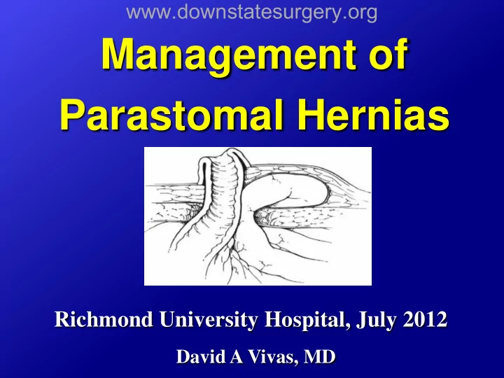
Management of Parastomal Hernias
Richmond University Hospital, July 2012
David A Vivas, MD
www.downstatesurgery.org
Management of Parastomal Hernias Richmond University Hospital, July - - PowerPoint PPT Presentation
www.downstatesurgery.org Management of Parastomal Hernias Richmond University Hospital, July 2012 David A Vivas, MD www.downstatesurgery.org Case Presentation HPI 85 y/o male s/p APR in 1978 for rectal cancer, no chemo/RT S/p TURP,
David A Vivas, MD
www.downstatesurgery.org
www.downstatesurgery.org
www.downstatesurgery.org
www.downstatesurgery.org
www.downstatesurgery.org
www.downstatesurgery.org
hernia
and lateral to the stoma in the LUQ
to the stoma
www.downstatesurgery.org
from surrounding tissues down to the level of the fascia
reduced
the ostomy.
www.downstatesurgery.org
interrupted #1 Prolene, extending both form the lateral and medial aspect of the hernia defect
www.downstatesurgery.org
www.downstatesurgery.org
www.downstatesurgery.org
www.downstatesurgery.org
www.downstatesurgery.org
www.downstatesurgery.org
www.downstatesurgery.org
www.downstatesurgery.org
www.downstatesurgery.org
www.downstatesurgery.org
www.downstatesurgery.org
www.downstatesurgery.org
www.downstatesurgery.org
www.downstatesurgery.org
www.downstatesurgery.org
www.downstatesurgery.org
www.downstatesurgery.org
www.downstatesurgery.org
www.downstatesurgery.org
www.downstatesurgery.org
www.downstatesurgery.org
www.downstatesurgery.org
www.downstatesurgery.org
www.downstatesurgery.org
www.downstatesurgery.org
www.downstatesurgery.org
www.downstatesurgery.org
www.downstatesurgery.org
Classification:
hernia space produced between the layers of the prolapsed bowel
alongside the bowel for the stoma
www.downstatesurgery.org
www.downstatesurgery.org
www.downstatesurgery.org
www.downstatesurgery.org
www.downstatesurgery.org
www.downstatesurgery.org
www.downstatesurgery.org
www.downstatesurgery.org
www.downstatesurgery.org
www.downstatesurgery.org
www.downstatesurgery.org
www.downstatesurgery.org
www.downstatesurgery.org
www.downstatesurgery.org
www.downstatesurgery.org
www.downstatesurgery.org
www.downstatesurgery.org
www.downstatesurgery.org
www.downstatesurgery.org
www.downstatesurgery.org
www.downstatesurgery.org
www.downstatesurgery.org
www.downstatesurgery.org
www.downstatesurgery.org
www.downstatesurgery.org
www.downstatesurgery.org
www.downstatesurgery.org
www.downstatesurgery.org
www.downstatesurgery.org
www.downstatesurgery.org
www.downstatesurgery.org
www.downstatesurgery.org
Strattice)
www.downstatesurgery.org
www.downstatesurgery.org
www.downstatesurgery.org
www.downstatesurgery.org
www.downstatesurgery.org
– The Sugarbaker technique had less recurrences than the keyhole technique (OR 2.3, 95% CI 1.2-4.6; p=0.016)
www.downstatesurgery.org
abandoned because of increased recurrence rates
significantly reduces recurrence rates and is safe with a low overall rate of mesh infection
superior over the keyhole technique showing fewer recurrences
www.downstatesurgery.org
www.downstatesurgery.org
www.downstatesurgery.org
www.downstatesurgery.org
www.downstatesurgery.org
www.downstatesurgery.org
reported
was less than 15% for all studies included
www.downstatesurgery.org
www.downstatesurgery.org
– To reduce the incidence of parastomal hernia by implanting a lightweight mesh in the sublay positions
www.downstatesurgery.org
group (above the peritoneum and the posterior rectus sheath
every 6 months after surgery
www.downstatesurgery.org
– Homogeneous groups (clinical and demographics) – Surgical time and postoperative morbidity similar in both groups – Zero mortality – No mesh intolerance
www.downstatesurgery.org
– Median 29 months (13-49) – 11/27 (40.7%) hernias in control group – 4/27 (14.8%) in study group – p=0.03
– 14/27 (44.4%) hernias in control group – 6/27 (22.2%) in study group
– p=0.08
www.downstatesurgery.org
– Parastomal placement of a mesh reduces the appearances of parastomal hernia – The technique is safe , well tolerated and does not increase morbidity rates
www.downstatesurgery.org
125
the Use of a Mesh to Prevent Parastomal Hernia. Ann Surg 2009;249:583-587
A Systematic Review of the Literature. Ann Surg 2012;255:685-695
http://www.fascrs.org
www.downstatesurgery.org
www.downstatesurgery.org
www.downstatesurgery.org
muscle
www.downstatesurgery.org
muscle
www.downstatesurgery.org
www.downstatesurgery.org
www.downstatesurgery.org
www.downstatesurgery.org
www.downstatesurgery.org
www.downstatesurgery.org