
SLIDE 1
1
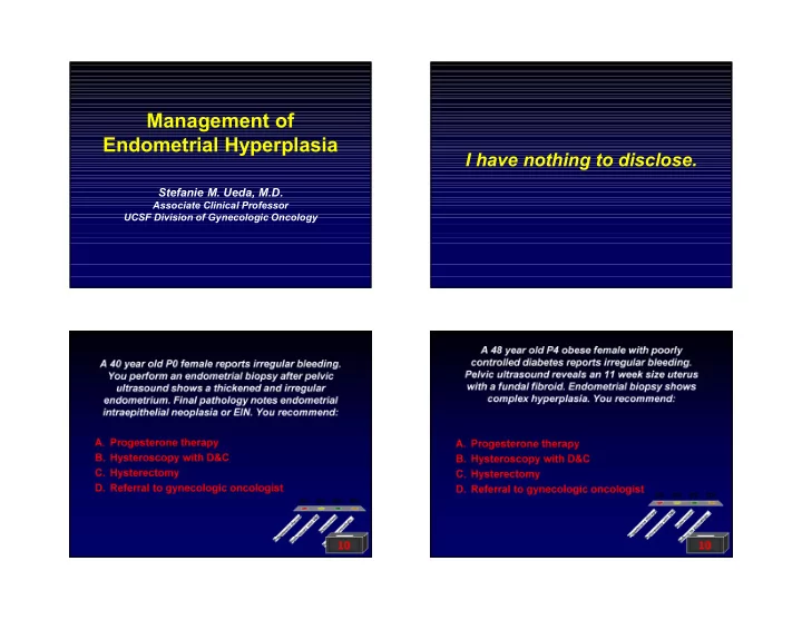
Management of Endometrial Hyperplasia
Stefanie M. Ueda, M.D.
Associate Clinical Professor UCSF Division of Gynecologic Oncology
I have nothing to disclose.
A 40 year old P0 female reports irregular bleeding. You perform an endometrial biopsy after pelvic ultrasound shows a thickened and irregular
- endometrium. Final pathology notes endometrial
intraepithelial neoplasia or EIN. You recommend:
- A. Progesterone therapy
- B. Hysteroscopy with D&C
- C. Hysterectomy
- D. Referral to gynecologic oncologist
0% 0% 0% 0%
10 A 48 year old P4 obese female with poorly controlled diabetes reports irregular bleeding. Pelvic ultrasound reveals an 11 week size uterus with a fundal fibroid. Endometrial biopsy shows complex hyperplasia. You recommend:
- A. Progesterone therapy
- B. Hysteroscopy with D&C
- C. Hysterectomy
- D. Referral to gynecologic oncologist
0% 0% 0% 0%
10