
SLIDE 1
5/23/2014 1
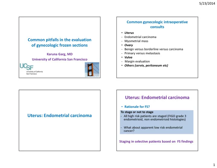
Common pitfalls in the evaluation
- f gynecologic frozen sections
Karuna Garg, MD University of California San Francisco
Common gynecologic intraoperative consults
- Uterus
- Endometrial carcinoma
- Myometrial mass
- Ovary
- Benign versus borderline versus carcinoma
- Primary versus metastasis
- Vulva
- Margin evaluation
- Others (cervix, peritoneum etc)
Uterus: Endometrial carcinoma Uterus: Endometrial carcinoma
- Rationale for FS?
To stage or not to stage
- All high risk patients are staged (FIGO grade 3
endometrioid, non endometrioid histologies)
- What about apparent low risk endometrial