Squamous Cell Carcinoma Squamous Cell Carcinoma of Precursors - - PowerPoint PPT Presentation

squamous cell carcinoma
SMART_READER_LITE
LIVE PREVIEW

Squamous Cell Carcinoma Squamous Cell Carcinoma of Precursors - - PowerPoint PPT Presentation

Squamous Cell Carcinoma Squamous Cell Carcinoma of Precursors Squamous intraepithelial lesions the Vulva and its Precursors Mimics Herpes infection, Syphilis Charles Zaloudek, M.D. Papillomatosis University of California, San


slide-1
SLIDE 1

1

Squamous Cell Carcinoma of the Vulva and its Precursors

Charles Zaloudek, M.D. University of California, San Francisco

Squamous Cell Carcinoma

  • Precursors

– Squamous intraepithelial lesions

  • Mimics

– Herpes infection, Syphilis – Papillomatosis – Multinucleated Atypia

  • Squamous cell carcinoma and variants

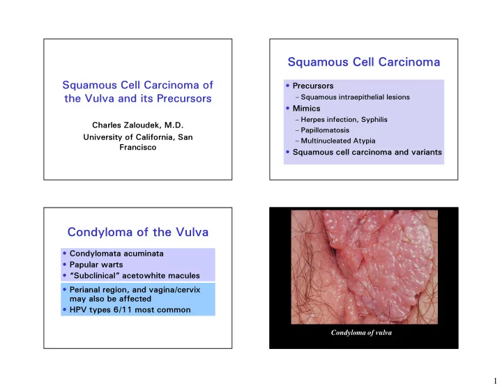
Condyloma of the Vulva

  • Condylomata acuminata
  • Papular warts
  • “Subclinical” acetowhite macules
  • Perianal region, and vagina/cervix

may also be affected

  • HPV types 6/11 most common

Condyloma of vulva

slide-2
SLIDE 2

2

Diagnostic Criteria for Condyloma of Vulva

  • Koilocytosis – cavitated cytoplasm
  • Nuclear atypia – enlargement, hyperchromasia,

pleomorphism, multinucleation, membrane irregularity

  • Architectural abnormalities – papillomatosis,

acanthosis, hyperkeratosis, parakeratosis

  • MIB-1 staining: Group of 2 or more stained

nuclei in same HPF in upper 2/3 of epithelium

  • More specific: HPV immunohistochemistry

and in situ hybridization

slide-3
SLIDE 3

3

Positive HPV Immunostain Positive HPV ISH

High Grade SIL (VIN)

Classic, or Bowenoid Type

  • Young women - 30’S and 40’s -

increasing in incidence

  • History of condylomas, herpes infection,

HIV disease, smoking

  • Linked to HPV, usually HPV 16
  • Varied appearance:

– Papules, plaques, polyps – White, red, or pigmented

  • Multicentric disease in vagina or cervix

Vulvar intraepithelial neoplasia Vulva - VIN III

slide-4
SLIDE 4

4

Pagetoid VIN

Pagetoid Lesions of the Vulva

HMWK LMWK CK 7 CEA S100 VIN

+

  • +
  • Melanoma
  • +

Paget’s

  • +

+ +

slide-5
SLIDE 5

5

High Grade SIL

Results of Therapy

  • Treatment: Excision, laser ablation,

topical imiquimod

  • Local recurrence - up to 35%
  • Occult invasion - up to 20%
  • Invasive carcinoma develops in

– 3-10% of treated patients – 9% to up to 90% of untreated patients – Invasive carcinoma is basaloid or condylomatous type, some keratinizing

  • Bowenoid papulosis

The Problem with Low Grade SIL (VIN)

  • Less than 50% of cases
  • f VIN I confirmed on

review

  • 70% of cases associated

with low risk HPV

  • MIB-1 positive nuclei in

upper epithelium correlate with VIN I

Logani S, et al. Mod Pathol 2003;16(8):735-741

Status of VIN Classification Pre-LAST

  • Category of VIN I eliminated

– ISSVD – WHO 2004

  • Most cases VIN I flat condyloma or

reactive

  • Diagnosis of VIN refers to high grade

lesions only

  • Classical and Differentiated types

Low Grade SIL of the Vulva

  • Some papillomatous with prominent

koilocytotic atypia (~ 1/3)

  • Some flat with minimal koilocytotic

atypia (~ 2/3)

  • No difference in HPV type distribution
  • 91% had detectable HPV

Srodon M, et al. Am J Surg Pathol 2007;30:1513-1518

slide-6
SLIDE 6

6

VIN 1 with extensive koilocytotic atypia found to have HPV 59 VIN 1 with minimal koilocytotic atypia found to have HPV 68 VIN 3 with HPV 16

Srodon M, et al. Am J Surg Pathol 2007;30:1513-1518

HPV Types in Low Grade SIL of Various Sites

HPV T ype Vulva Vagina Cervix Low risk 67% 35% 6% 6 or 1 1 42% 6% High risk 42% 76% 94% 16 6% 6% 1 1% 18 24% 17%

Srodon M, et al. Am J Surg Pathol 2007;30:1513-1518

HPV Types in High Grade SIL of Various Sites

HPV T ype Vulva Vagina Cervix Low risk 3% 6% 0% 6 or 1 1 3% 0% High risk 100% 94% 100% 16 91% 50% 75% 18 6 0% 4%

Srodon M, et al. Am J Surg Pathol 2007;30:1513-1518

VIN

Simplex or Differentiated Type

  • 2-10% of VIN
  • Postmenopausal women, average age late

60’s

  • Small lesions, roughened gray white, or

white plaques, may be multifocal

  • Lichen sclerosis may also be present
  • Not associated with HPV
  • ? Greater potential for progression to

invasive carcinoma; invasive carcinoma is keratinizing squamous cell type

slide-7
SLIDE 7

7

p53

slide-8
SLIDE 8

8

p53 MIB1

p53

slide-9
SLIDE 9

9

Differentiated vulvar intraepithelial neoplasia contains TP53 mutations and is genetically linked to vulvar squamous cell carcinoma.

Pinto AP, et al. Mod Pathol (2010) 23: 404-412.

  • 6 of 10 dVIN cases had at least 1 VIN focus

with 1 or more TP53 mutations

  • 4 were p53 immunopositive (missense or splice)

and 2 were p53 negative (deletions)

  • 5 had SCC associated with dVIN, 4/5 had TP53

mutations

– 2 missense, p53 positive – 2 nonsense, p53 negative

  • 2/4 cases had same TP53 mutation in dVIN and

SCC

  • Multiple foci dVIN with different mutations in

some cases

Expanding the morphologic spectrum of differentiated VIN (dVIN) through detailed mapping of cases with p53 loss. Singh N, et al. Am J Surg Pathol, 2015;39:52-60

  • p53 negative dVIN and SCC, when present,

in 14 specimens from 10 patients

  • 27% of dVIN cases at Vancouver General

Hospital

  • 1 case reclassified as not dVIN
  • In 5/13 the p53 negative areas corresponded

to the morphologic dVIN

  • In 8/13 the p53 negative areas were more

extensive than the morphologic dVIN and in some were at margins

Expanding the morphologic spectrum of differentiated VIN (dVIN) through detailed mapping of cases with p53 loss. Singh N et al. Am J Surg Pathol 2015;39:52-60

slide-10
SLIDE 10

10 p53 CK17

FIGURE 1. A, Simplex vulvar intraepithelial neoplasia, grade 3 of basaloid type. Diffuse replacement of the whole epidermis by a homogeneous population of small, "undifferentiated" keratinocytes with scanty cytoplasm extending throughout the entire thickness of the epidermis, showing no or only minimal maturation in superficial layers. Normal vulvar squamous epithelium is present on the left. B, The epidermis is thickened and shows a parakeratotic surface reaction. The rete ridges are elongated and markedly enlarged and coalescent. C, Small cells, with scant cytoplasm, showing large vesicular nuclei, with visible nucleoli. D, Moderate to severe atypia, with nuclear pleomorphism, multinucleation, and dyskeratosis.

Ordi J et al. Am J Surg Pathol 2009 33:1659-1665

p16

slide-11
SLIDE 11

11

LAST Project

Lower Anogenital Squamous Terminology Standardization Project for HPV-associated Lesions

  • Single set of diagnostic terms for all lower

anogenital sites

  • Two-tiered nomenclature for intraepithelial

lesions

– Low grade generally self-limited lesions – High grade potentially progressive lesions

  • Recommended terminology:

– Low grade SIL – High grade SIL

  • Use p16 only to improve accuracy
  • Vulvar SISCCA = FIGO IA cancer

Int J Gynecol Pathol 2013; 32:76-1 15

Current Classification of Vulvar Squamous Intraepithelial Lesions

WHO, 2014 Low grade SIL High grade SIL Differentiated type VIN ISSVD, 2015 Low grade SIL (vulvar LSIL, flat condyloma or HPV effect) High grade SIL (vulvar HSIL, VIN usual type) Differentiated type VIN

  • Bornstein J, et al. Obstet Gynecol

2016;127:264-268

  • Sideri M, et al. Am J Surg Pathol

2007;31:1452

  • WHO Classification of

Tumours of the Female Reproductive Organs, 4th Ed, 2014

  • Stoler MH and Kurman RJ.

Am J Surg Pathol 2007;31;1452-1454

Squamous Cell Carcinoma

  • f the Vulva
  • Type I

– Younger women, average age 55 – VIN, HPV associated, usually HPV 16 – Basaloid, condylomatous types

  • Type II

– Older women, average age 77 – No or simplex VIN, no HPV, some LS or SH – Keratinizing squamous cell carcinoma

slide-12
SLIDE 12

12

HPV and Squamous Cell Carcinoma of the Vulva

  • HPV prevalence in vulvar

intraepithelial neoplasia: 52% to 100%

  • HPV prevalence in invasive

squamous cell carcinoma: 15% to 79%

Histopathology 2013; 62:161-175

HPV Testing in Vulvar Carcinoma

Toki, Kurman, et al 1991 Ave. Age HPV+ Adj. VIN Adj. SH, LS Adj Nl SCC 77 4/19 2/19 14/19 3/19 BC 54 6/8 7/8 1/8 WC 47 3/3 3/3 Ave. Age HPV+ Adj. VIN Adj. SH, LS Adj Nl SCC 77 4/19 2/19 14/19 3/19 BC 54 6/8 7/8 1/8 WC 47 3/3 3/3

VIN and LS adjacent to thin vulvar SCC

T ype Ca Classic VIN Differentiated VIN No VIN Keratinizing SCC-38 9 18 1 1 Warty SCC-6 6 Basaloid SCC-4 4 LS 1/19 9/16 4/9 Age Pt. 62 78 75

Am J Surg Pathol 2006;30:310-318

HPV in SCC of the Vulva

  • Worldwide study of 2296 cases, 587

VIN and 1709 invasive SCC

– 25.1% SCC were HPV related – Prevalence of HPV related highest in young women – Basaloid and warty most likely to be HPV positive – HPV 16 most common (72.5%, followed by HPV 33 (6.5%) and HPV 18 (4.6%)

De Sanjose, S, et al. Eur J Cancer 2013;49:3450-3461

slide-13
SLIDE 13

13

Squamous Cell Carcinoma

  • f the Vulva
  • Most occur on labia
  • Clitoris involved in

<15%

  • Multifocal in <10%
  • Exophytic papillary

mass or endophytic ulcer

Grading of Vulvar Squamous Cell Carcinoma

  • Grade 1: No poorly

differentiated component

  • Grade 2: < 50%

poorly differentiated component

  • Grade 3: > 50%

poorly differentiated component

1 2 3 1

Squamous Cell Carcinoma of the Vulva

Subtype Keratinizing Non-keratinizing Basaloid Warty (Condylomatous) Verrucous

slide-14
SLIDE 14

14

Basaloid T ype of Squamous Cell Carcinoma

slide-15
SLIDE 15

15

Condylomatous T ype of Squamous Cell Carcinoma

Squamous Cell Carcinoma of the Vulva Therapy and Results

  • Radical vulvectomy in the past
  • Treatment now more conservative -

wide excision, hemivulvectomy for lateralized tumors

  • About 30% of patients have LN

metastases, lymphadenectomy generally performed; role for SLN Bx

  • Survival 75% overall, 90-100% stage I
  • The term “microinvasive carcinoma” is not used

in the vulva

  • The category of stage IA is an attempt to define

a group with a very low risk of lymph node metastasis

  • Definition: ≤ 1 mm in depth and ≤ 2 cm diameter
  • Lymphovascular invasion, growth pattern do not

exclude tumors from this category

Superficially Invasive Squamous Cell Carcinoma

  • f the Vulva (Stage IA)

Histologic Features Suggestive

  • f Invasion

Rete ridges are irregular in size, shape and distribution Rete ridges extend deeply into the dermis Complex budding or branching of the rete ridges Paradoxical keratinization deep in the rete ridges or in dermal nests Prong like buds of epithelium grow into the dermis Irregular, often angulated nests of atypical squamous cells in the dermis T

  • o many (crowded) nests of cells, often irregularly distributed

Single or small clusters of atypical cells in the dermis Cells in the dermis have vesicular nuclei, prominent nucleoli, eosinophilic cytoplasm – different from adjacent VIN Desmoplastic or edematous stroma around dermal nests Growth adjacent to or around thick walled blood vessels or nerves

slide-16
SLIDE 16

16 Squamous Cell Carcinoma of the Vulva Measurements of Invasion

Depth = From the epithelial- stromal junction of the adjacent most superficial dermal papilla to the deepest point of invasion Depth Thickness

Verrucous Carcinoma of the Vulva

  • Condyloma misclassified as verrucous

carcinoma

  • Verrucous carcinoma misclassified as

carcinoma

  • Well differentiated SCC misclassified as

verrucous carcinoma

  • Verrucous carcinoma misclassified as

well differentiated SCC

slide-17
SLIDE 17

17

Verrucous Carcinoma of the Vulva

  • Elderly women
  • Locally aggressive with eventual

formation of a large warty tumor

  • Lymph node metastases are rare
  • Treatment is by wide local excision;

vulvectomy may be necessary

  • 5-year survival ~ 80%

Findings Adjacent to Verrucous Carcinoma of Vulva

  • Lichen Sclerosis – 1
  • LSC with verrucous features – 7
  • VAAD – 7

– Vulvar acanthosis with altered differentiation – Variable verruciform architecture – Plaque like parakeratosis – Cytoplasmic pallor due to loss of granular layer

  • No Classic or differentiated VIN

Am J Surg Pathol 2004;28:638-643

Vulvar acanthosis with altered differentiation

Am J Surg Pathol 2004;28:638-643

S = hyperplasia, sharp B = hyperplasia, blunt C = verrucous carcinoma

VAAD

slide-18
SLIDE 18

18

Despite the name “verrucous” carcinoma… Most think that there is no good evidence of an association with HPV

slide-19
SLIDE 19

19

Where Does This Information Come From?

  • Edward J.

Wilkinson, MD

  • “Verrucous

carcinoma may be associated with HPV, typically type 6 or variants of type 6.”

Verrucous Carcinoma and HPV

  • 27 cases initially classified as VC reviewed; after

review 13 accepted

  • 11 cases initially classified as VC of

vulva/perineum

– 5 accepted as VC – none had HPV – 4 reclassified as SCC – none had HPV – 2 reclassified as giant condylomas, 1 had HPV 6, 1 had HPV 11

  • Conclusions

– HPV unlikely to be causally related to VC – Positive HPV test favors giant condyloma over VC

Mod Pathol 2012; 25:1354-1363

Verrucous Carcinoma Large/Giant Condyloma ≠ Low risk HPV in situ in a giant condyloma

slide-20
SLIDE 20

20

Differences Between Verrucous Carcinoma and Condyloma

  • Verrucous carcinoma is usually large,

condyloma is smaller

  • Verrucous carcinoma shows pushing

invasion into the underlying dermis, condyloma grows off the surface

  • Usually more abnormal cytology in verrucous

carcinoma

  • No koilocytosis in verrucous carcinoma,

characteristic of condyloma

  • No HPV in verrucous carcinoma, HPV

present in condyloma

Not verrucous carcinoma; well differentiated squamous cell carcinoma Not verrucous carcinoma!

slide-21
SLIDE 21

21

“Pseudotumors” that Mimic Squamous Cell Carcinoma

Case 1

  • 37 year old HIV positive woman
  • Vulvar tumor excised
slide-22
SLIDE 22

22

Chronic hypertrophic vulvar herpes simulating neoplasia. International Journal of Gynecological Pathology 2012;31(1):33-37.

We present a case of a 40-year-old woman with a history of human immunodeficiency virus infection and a nodular, hyperkeratotic 3.5-cm vulvar mass that increased in size over a 2-month period. Histopathologic examination of the excised mass was diagnostic of chronic hypertrophic vulvar herpes simulating neoplasia. Hypertrophic vulvar herpes presents a diagnostic challenge for both pathologists and clinicians because of its unusual clinicopathologic features that mimic neoplasia and its rarity. There is therefore the need for the correct diagnosis of this entity, so that appropriate therapy can be given. The pertinent literature is reviewed and discussed.

Case 2

  • 39 years old, HIV positive
  • Clinical diagnosis: Malignant

neoplasm

  • 3 cm fragment of mucosal tissue

excised

  • Histology: ? Squamous cell carcinoma
slide-23
SLIDE 23

23

Histologic Features of Condyloma Latum

  • Acanthosis
  • Elongation and broadening of rete

pegs

  • Neutrophil infiltration of the epidermis
  • Mononuclear infiltration of the dermis
  • Spirochetes in zone of PMN infiltrate

Freinkel AL Histopathology 1987, 1 1:819-831

Paget Disease of the Vulva

56 Cases Studied at Duke

  • Mean age at diagnosis 69
  • Pruritus, erythematous white plaque
  • Average size 5.6 cm, 54% unilateral
  • 68% had no recurrence
  • 68% of curative resections had + margins
  • Invasive Paget’s in 18%, 0.2-6mm
  • Last seen: 43% NED, 43% DOC, 9% AwP,

3% lost, 2% (only 1 patient) DIP

International Journal of Gynecological Pathology. 29(1):69-78, January 2010.

slide-24
SLIDE 24

24

Paget’s Disease

Use of Immunohistochemistry

Stain Vulvar No Assoc Ca Vulvar Assoc Ca Perianal No Assoc Ca Perianal Assoc Ca LMWK

+ + + +

CEA

+ + + +

CK7

+ + + +

CK20

  • +/-
  • +

GCDFP

+

  • +
  • Vulvar Paget’s Disease

CK20 CK7 GCDFP-15

CK20+, CDX-2+ and GCDFP -

Paget’s Disease is likely secondary