Case 1 Illuminating Consultation 44 year old woman with a mass in - - PowerPoint PPT Presentation

case 1
SMART_READER_LITE
LIVE PREVIEW

Case 1 Illuminating Consultation 44 year old woman with a mass in - - PowerPoint PPT Presentation

5/25/2013 Case 1 Illuminating Consultation 44 year old woman with a mass in the Cases rectus muscle. Biopsy performed, reviewed at UCSF. Immunostains evaluated: Charles Zaloudek, M.D. Positive: Pankeratin, keratin AE1/AE3, CK7


slide-1
SLIDE 1

5/25/2013 1

Illuminating Consultation Cases

Charles Zaloudek, M.D. Department of Pathology University of California San Francisco
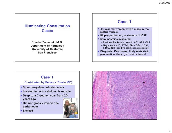
Case 1

  • 44 year old woman with a mass in the

rectus muscle.

  • Biopsy performed, reviewed at UCSF.
  • Immunostains evaluated:

– Positive: Pankeratin, keratin AE1/AE3, CK7 – Negative: CK20, TTF-1, ER, CD34, CD31, S100, INI1 (positive stain, negative result)

  • Diagnosis: Carcinoma, likely metastatic,

pancreaticobiliary, gyn, skin adnexal

Case 1

(Contributed by Rebecca Swain MD)

  • 9 cm tan-yellow whorled mass
  • Located in rectus abdominis muscle
  • Deep to a C-section scar from 20

years ago

  • Did not grossly involve the

peritoneum

  • Excised
slide-2
SLIDE 2

5/25/2013 2 Tumors and Masses of the Abdominal Wall

(From Current Diagnosis and Treatment: Surgery)

  • Hernia
  • Lipoma
  • Hemangioma
  • Endometriosis
  • Fibromatosis (desmoid tumor)
  • Malignant tumor – most are

metastatic, lung and pancreas are the most common primary sites

slide-3
SLIDE 3

5/25/2013 3

Summary

  • Young woman
  • Tumor in the anterior abdominal wall
  • Pan keratin and CK7 positive
  • No internal primary site identified
  • Related to a C-section scar
  • PO Day 1 serum hCG = 27 IU/L, PO

Day 3 serum hCG = 9 IU/L

Immunohistochemistry of Trophoblastic Tumors

Best Marker MIB1 Rate Choriocarcinoma hCG +++ ETT p63 + or ++ PSTT hPL + or ++

General trophoblastic markers: HSD3B1, CD10, inhibin, CK18, pankeratin

Inhibin

Case 1 Another Case

HCG HPL p63

slide-4
SLIDE 4

5/25/2013 4

Ki-67

Diagnosis Epithelioid Trophoblastic Tumor

Description of 14 Cases of A New Type of Trophoblastic Tumor

  • Shih IM and Kurman RJ. Epithelioid

trophoblastic tumor – A neoplasm distinct from choriocarcinoma and placental site trophoblastic tumor simulating carcinoma. Am J Surg Pathol, 1998; 22:1393-1403.

Epithelioid Trophoblastic Tumor

  • Patient age 15-66 (average 36)
  • Antecedent pregnancy:

– Normal delivery 68% – Spontaneous abortion 16% – Hydatidiform mole 16%

  • Interval 1-25y (average 6.2)
  • Presentation: amenorrhea or abnormal

bleeding – can be misdiagnosed as ectopic

  • Some patients present with extrauterine

tumors – lung or other sites

  • Low levels of HCG
slide-5
SLIDE 5

5/25/2013 5

Epithelioid Trophoblastic Tumor

  • DNA genotyping reveals paternal

alleles that are not present in adjacent maternal tissues trophoblastic

  • rigin
  • Conflicting data on Y-chromosome,

but a recent study suggested that most ETT do not have Y chromosome material present (paternal X favored)

p63 hPL

Chorionic IT – Chorion Laeve

Epithelioid T rophoblastic T umor Cervix

ETT Location of Primary Tumor

  • Cervix or LUS

50%

  • Corpus

30%

  • Extrauterine

20%

  • Moral: Can be easily mistaken for

cervical cancer

slide-6
SLIDE 6

5/25/2013 6

Coulson, L. E., et al. (2000). "Epithelioid trophoblastic tumor of the uterus in a postmenopausal woman - A case report and review of the literature." American Journal of Surgical Pathology 24(1 1): 1558-1562.

slide-7
SLIDE 7

5/25/2013 7

ETT - CK18 HPL HCG ETT Immunohistochemistry:

  • Scattered HPL, HCG, PLAP

, Mel-CAM + cells.

  • p63+, HLA-G+
  • MIB1 10-25%

ETT

One of the original descriptions

Mazur MT. Metastatic gestational choriocarcinoma. Unusual pathologic variant following

  • chemotherapy. Cancer.

1989; 63:1370-1377. Two lung tumors reported.

slide-8
SLIDE 8

5/25/2013 8

p63 ETT in the Lung CK18

Epithelioid Trophoblastic Tumor

  • Clinical behavior similar to PSTT
  • Most have benign clinical evolution
  • ~25% metastasize
  • Serum HCG, although low, can be

used to monitor treatment

  • May not respond to standard GTD

chemotherapy

  • Hysterectomy is standard therapy
  • Resection of metastases may help

Case 1 Key Points

  • ETT is derived from intermediate trophoblasts of the

chorion laeve

  • Low levels of serum HCG
  • Usually primary in the cervix or uterus
  • Nodular, epithelioid appearance resembles a carcinoma
  • Differential diagnosis

– Squamous cell carcinoma of the cervix: CK5/6, p16, HPV testing; LMW CK, staining for trophoblastic markers, molecular genotyping – Placental site nodule: size, cellularity, low MI, low Ki-67

  • Difficult to treat with chemotherapy, hysterectomy is

usually performed

  • Extrauterine primary tumors occur in the lungs, pelvis

and elsewhere

Gestational Trophoblastic Neoplasia

  • Persistent or invasive hydatidiform

mole

  • Choriocarcinoma
  • Epithelioid Trophoblastic Tumor (ETT)
  • Placental Site Trophoblastic Tumor

(PSTT)

  • Mixed Trophoblastic Tumor
slide-9
SLIDE 9

5/25/2013 9

Case 2

(Contributed by Dave Park)

  • 41 year old G5P3
  • Persistently elevated HCG in the

range of 200-300 IU/L

  • Underwent a D&C for

suspected missed abortion.

slide-10
SLIDE 10

5/25/2013 10

Desmin hCG p63 hPL

Immunohistochemistry of Trophoblastic Tumors

Best Marker MIB1 Rate Choriocarcinoma hCG +++ ETT p63 + or ++ PSTT hPL + or ++

General trophoblastic markers: HSD3B1, CD10, inhibin, CK18, pankeratin

Summary

  • Intrauterine
  • Ill defined, invades the myometrium
  • Polygonal cells, atypia, mitotic

activity

  • Low HCG and p63, extensive HPL
  • Diagnosis: Placental site trophoblastic

tumor (PSTT)

slide-11
SLIDE 11

5/25/2013 11

Trophoblastic Pseudotumor of the Uterus

An exaggerated form of “Syncytial Endometritis” Simulating a Malignant Tumor Robert J Kurman, MD (MAJ, MC, USA), Robert E Scully, MD, and Henry J Norris, MD Cancer 38:1214-1226, 1976

  • 1. Twiggs LB, Okagaki T, Phillips GL, Stroemer JR, Adcock
  • LL. Trophoblastic pseudotumor-evidence of malignant

disease potential. Gynecologic Oncology 1981;12:238-48.

  • 2. Gloor E, Dialdas J, Hurlimann J, Ribolzi J, Barrelet L.

Placental site trophoblastic tumor (trophoblastic pseudotumor) of the uterus with metastases and fatal

  • utcome. Clinical and autopsy observations of a case.

American Journal of Surgical Pathology 1983;7:483-6.

  • 3. Eckstein RP, Russell P, Friedlander ML, Tattersall MH,

Bradfield A. Metastasizing placental site trophoblastic tumor: a case study. Human Pathology 1985;16:632-6.

Scully RE, Y

  • ung RH. T

rophoblastic pseudotumor: A reappraisal. Am J Surg Pathol 1981; 5:75-76. Proposed name Placental Site T rophoblastic T umor

Placental Site Trophoblastic Tumor PSTT

  • Mean age about 31y, range 20 to 63y
  • 60-70% follow an uneventful term pregnancy,

the rest follow CHM or abortion

  • Requirement for a paternal X chromosome;

antecedent gestation female

  • Time from previous pregnancy to PSTT

diagnosis 1 week to 204m, median 12-18m

  • Presentation is with vaginal bleeding, uterine

enlargement

  • Usually slight to moderate elevation of serum

hCG

  • About 85% of cases are in stage I at diagnosis

PSTT at NETDC

(Gynecol Oncol 82:415-419, 2001)

  • 13 patients, average age lower 30’s
  • Most antecedent pregnancies normal or

abortion; hydatiform mole rare

  • Average time from antecedent pregnancy to

diagnosis ~16m (2w - 5y)

  • Serum hCG slightly elevated (< 500

mIU/ml)

  • Treatment usually by hysterectomy; 4 had

pelvic metastases at diagnosis

  • 43% had recurrences; all had received

chemotherapy

slide-12
SLIDE 12

5/25/2013 12

Placental Site T rophoblastic T umor – Gross Appearance A tumor mass is almost always present Ill defined edge, not sharply circumscribed

slide-13
SLIDE 13

5/25/2013 13

slide-14
SLIDE 14

5/25/2013 14

PSTT Immunohistochemistry

  • Positive

– Cytokeratin cocktail – LMW cytokeratin (CK18) – Inhibin – hPL (97% +, 56% strong +) – CD146 (MelCAM), HLA-G

  • Negative or weak

– hCG (almost always scattered + cells) – PLAP, p63

PSTT - CK18

HCG HPL

PSTT - MIB1

slide-15
SLIDE 15

5/25/2013 15

Differential Diagnosis

  • Squamous cell carcinoma: Different

appearance, different immunophenotype

  • Choriocarcinoma: Different cell

population, less proliferation, different HCG profile, different immunophenotype

  • ETT: Related, but different cytology,

different low power appearance, different immunophenotype

Differential Diagnosis

  • Exaggerated placental site reaction

– A benign expansion of the normal population of implantation site intermediate trophoblasts in the placental bed – High cellularity can mimic PSTT – No mass in EPS Reaction – Can be very cellular, but lacks cohesive masses of cells – Does not permeate and splay the myometrium – Minimal mitotic activity and Ki-67 immunoreactivity – Lack of a genetic association with PSTT – majority have a paternal Y chromosome

EPS CK hPL Ki-67

Exaggerated Placental Site Reaction

slide-16
SLIDE 16

5/25/2013 16

PSTT Clues to the Diagnosis

  • Low levels of HCG
  • Tumor mass present – check the ultrasound
  • r other imaging
  • Sheets of implantation site intermediate

trophoblasts

  • Invasion of myometrium by nests and sheets
  • f intermediate trophoblasts
  • Mitotic figures and increased MIB1 (~10%)
  • HPL + many cells, HCG + few cells
  • No chorionic villi

PSTT Behavior

  • ~ 15% clinically malignant
  • Primary treatment is surgical
  • Response to chemotherapy variable

– EMA/CO 71% respond, 38% have a complete response

  • Unfavorable prognostic factors

– Stage > I (extrauterine spread) – Mitotic index >5 mf/10 hpf – Large tumor size, clear cells – Interval from antecedent pregnancy to diagnosis (> 2 y less favorable)

Case 2 Key Points

  • PSTT is derived from implantation site

trophoblastic cells

  • Cohesive groups of IT form a mass and

invade the myometrium, splaying the muscle bundles

  • Intravascular tumor common
  • No villi
  • At least some mitotic activity
  • hPL, Ki-67 are the most useful immunostains

Cytotrophoblast Syncytiotrophoblast Villous IT in trophoblastic columns Implantation site IT p63-, HPL +++ Chorionic-type IT p63+++, HPL +/- Exaggerated placental site Placental site trophoblastic tumor Placental site nodule Epithelioid trophoblastic tumor Choriocarcinoma

slide-17
SLIDE 17

5/25/2013 17

Table 4. Immunohistochemical features of trophoblastic lesions of the uterus.

CK18 HLAG Inhibin HCG HPL p63 PLAP MIB1 PSN

  • +/- +/-
  • 5%<

ETT

  • +/- +/-

+/-

20% EPS

  • +/-
  • +/-

Near 0% PSTT

  • +/-
  • +/-

10% Chorio Ca

  • +/- +/-

+/-

> 50% Immunohistochemistry of T rophoblastic Proliferations

Case 3

  • Sessile vulvar lesion from a 66 year
  • ld female

– Is it verrucous carcinoma? – Is it invasive? – If it is invasive how deeply invasive is it? – Is further excision necessary? – Is lymph node sampling necessary?

slide-18
SLIDE 18

5/25/2013 18

Is it Verrucous Carcinoma?

What do you think?

What is Verrucous Carcinoma of the Vulva?

  • A very well differentiated variant of

squamous cell carcinoma

  • Occurs in elderly women
  • Locally aggressive with eventual formation
  • f a large warty tumor
  • Lymph node metastases are rare
  • Treatment is by wide local excision;

vulvectomy may be necessary, does not require lymph node dissection

  • 5-year survival - unknown
slide-19
SLIDE 19

5/25/2013 19

Diagnostic Criteria for Verrucous Carcinoma

  • Marked acanthosis, verrucous surface

contour

  • Large rounded bulbous rete ridges

push into the underlying tissue

  • Minimal pleomorphism
  • Coarse chromatin, variable nucleoli
  • Infrequent mitotic figures
  • No koilocytosis
slide-20
SLIDE 20

5/25/2013 20

S = hyperplasia, sharp B = hyperplasia, blunt C = verrucous carcinoma Not verrucous carcinoma! Our case: Not verrucous carcinoma; well differentiated squamous cell carcinoma S = hyperplasia, sharp B = hyperplasia, blunt C = verrucous carcinoma

slide-21
SLIDE 21

5/25/2013 21

Vulvar acanthosis with altered differentiation

Am J Surg Pathol 2004;28:638-643

“VAAD”

Despite the name “verrucous” carcinoma… No good evidence of an association with HPV Verrucous Carcinoma and HPV

  • 27 cases initially classified as VC reviewed;

after review 13 accepted

  • 11 cases initially classified as VC of

vulva/perineum

– 5 accepted as VC – none had HPV – 4 reclassified as SCC – none had HPV – 2 reclassified as giant condylomas, 1 had HPV 6, 1 had HPV 11

  • Conclusions

– HPV unlikely to be causally related to VC – Positive HPV test favors giant condyloma over VC

Mod Pathol 2012; 25:1354-1363

slide-22
SLIDE 22

5/25/2013 22

Differences Between Verrucous Carcinoma and Condyloma

  • Verrucous carcinoma is usually large,

condyloma is smaller

  • Verrucous carcinoma shows pushing

invasion into the underlying dermis, condyloma grows off the surface

  • Usually more abnormal cytology in verrucous

carcinoma

  • No koilocytosis in verrucous carcinoma,

characteristic of condyloma

  • No HPV in verrucous carcinoma, HPV

present in condyloma

Condyloma Verrucous Carcinoma Verrucous Carcinoma Condyloma

slide-23
SLIDE 23

5/25/2013 23

Positive HPV Immunostain

Classification of Vulvar Squamous Cell Carcinoma

T ype HPV Associated Frequency Keratinizing SCC Some, most HPV

  • Most common

Basaloid SCC Y es Occasional Warty or condylomatous SCC Y es Occasional Verrucous SCC No Rare

VIN adjacent to thin vulvar SCC

T ype Ca Classic VIN Differentiated VIN No VIN Keratinizing SCC-38 9 18 1 1 Warty SCC-6 6 Basaloid SCC-4 4 Age Pt. 62 78 75

Am J Surg Pathol 2006;30:310-318

More Questions?

  • Is it invasive?
  • What is the depth?
  • Does the patient need more

treatment?

slide-24
SLIDE 24

5/25/2013 24

Histologic Features Suggestive

  • f Invasion

Rete ridges are irregular in size, shape and distribution Rete ridges extend deeply into the dermis Complex budding or branching of the rete ridges Paradoxical keratinization deep in the rete ridges or in dermal nests Prong like buds of epithelium grow into the dermis Irregular, often angulated nests of atypical squamous cells in the dermis T

  • o many (crowded) nests of cells, often irregularly distributed

Single or small clusters of atypical cells in the dermis Cells in the dermis have vesicular nuclei, prominent nucleoli, eosinophilic cytoplasm – different from adjacent VIN Desmoplastic or edematous stroma around dermal nests Growth adjacent to or around thick walled blood vessels or nerves

Squamous Cell Carcinoma of the Vulva Measurements of Invasion

Depth = From the epithelial- stromal junction of the adjacent most superficial dermal papilla to the deepest point of invasion

  • The term “microinvasive carcinoma” is not used

in the vulva

  • The category of stage IA is an attempt to define

a group with a very low risk of lymph node metastasis

  • Definition: ≤ 1 mm in depth and ≤ 2 cm diameter
  • Lymphovascular invasion, growth pattern do not

exclude tumors from this category

Superficially Invasive Squamous Cell Carcinoma

  • f the Vulva (Stage IA)
slide-25
SLIDE 25

5/25/2013 25

Depth Thickness Depth of Invasion = 6.5 mm Width = 13 mm Length = 21 mm 1 mm from the deep margin

Staging of Vulvar Squamous Cell Carcinoma

Location Size Depth Stage Ia Confined to vulva 2 cm or less 1 mm or less Stage Ib Confined to vulva > 2 cm Any tumor > 1 mm, regardless

  • f diameter
slide-26
SLIDE 26

5/25/2013 26

Our Case

  • Stage Ib
  • More treatment required
  • Lymph node dissection or, possibly,

sentinel lymph node biopsy

  • ? Re-excision to get a wider margin

Squamous Cell Carcinoma of the Vulva Therapy and Results

  • Radical vulvectomy in the past
  • Treatment now more conservative -

wide excision, hemivulvectomy for lateralized tumors

  • About 30% of patients have LN

metastases, lymphadenectomy generally performed; role for SLN Bx

  • Survival 75% overall, 90-100% stage I

Case 3 Key Points

  • Must meet specific criteria to be diagnosed

as verrucous carcinoma: very well differentiated, pushing invasion, rounded rete ridges

  • Prongs, irregular nests, infiltrative patterns,

higher grade exclude verrucous carcinoma regardless of the surface contour

  • Depth of invasion and size are critical
  • Any tumor that exceeds 1 mm in depth is

likely to be treated with lymph node dissection

  • Only stage Ia is treated conservatively