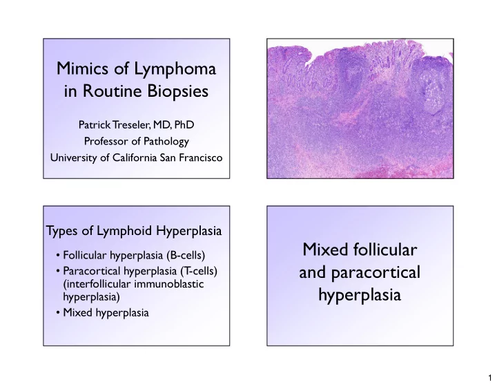

Mimics of Lymphoma in Routine Biopsies Patrick Treseler, MD, PhD Professor of Pathology University of California San Francisco Types of Lymphoid Hyperplasia Mixed follicular • Follicular hyperplasia (B-cells) • Paracortical hyperplasia (T -cells) and paracortical (interfollicular immunoblastic hyperplasia hyperplasia) • Mixed hyperplasia 1
Follicular Hyperplasia Mixed Follicular Hyperplasia and Paracortical Hyperplasia CD3 CD20 2
CD20 3
CD20 The “Panel o’ Three” (for assessment of lymphoid infiltrates) � CD20 � CD3 � CD21 CD21 CD3 4
CD20 CD20 5
CD20 CD20 6
CD3 CD21 Small B-Cell Lymphomas The “Panel o’ Nine” (for diagnosis of small B-cell lymphomas) Basic Immunophenotypes � CD20 � CD3 CD20 CD5 CD43 CD23 BCL1 BCL6 CD10 Cyclin D1 � CD5 CLL/SLL + + + + - - - � CD43 Mantle cell + + + - + - - � CD10 � CD21 Follicular + - - -/+ - + +/- � CD23 Marginal + - -/+ - - - - � BCL-1 (cyclin D1) � BCL-6 Proportion of cases positive: + >90%, +/- 50-90%, -/+ 10-50%, - <10% 7
Paracortical hyperplasia Differential diagnosis Paracortical • Drug reaction • Other hypersensitivity reaction hyperplasia • Viral infection • Post-vaccination • No clear etiology CD3 8
Florid paracortical hyperplasia CD20 (e.g., infectious mononucleosis) can mimic: • Diffuse large B-cell lymphoma, NOS • Classical Hodgkin lymphoma • T -cell/histiocyte-rich large B-cell lymphoma • EBV+ diffuse large B-cell lymphoma of the elderly • Peripheral T -cell lymphoma Infectious Mono Infectious Mono 9
CD20 Infectious Mono Infectious Mono CD3 “One should think twice and thrice before rendering a diagnosis of DLBCL in a patient younger than 20 years. Infectious mononucleosis in particular has to be suspected when … there are many admixed large T-cells and Waldeyer’s ring is involved.” ACL Chan & JKC Chan, 2011 Diffuse large B-cell lymphoma, in Hematopathology (Saunders/Elsevier) Infectious Mono 10
Infectious Mono EBV-ISH Slide courtesy of Dr. Dan Arber, Stanford Univ. Infectious-Mono-EBER-1000X CD30 CD20 Infectious Mono Infectious Mono 11
Classical Hodgkin lymphoma CD20 (CHL) HRS Cell Immunophenotype (Basic Panel for Dx of CHL) � CD30 + >90% � CD15 +/- ~80% � CD20 -/+ ~20% (focal, weak)* � Oct2 - ~60% (focal, weak)* � Pax-5 + >90% (often focal, weak)* � CD3 - <10% Proportion of cases positive: + >90%, +/- 50-90%, -/+ 10-50%, - <10% *Based on data from García-Cosío et al. Mod Pathol 17: 1531; 2004 CD20 T-cell/histiocyte-rich large BCL 12
T -cell/histiocyte-rich large BCL T -cell/histiocyte-rich large BCL Diagnostic criteria (WHO 2008) Boudová et al. (Blood 102: 3753; 2003) • Large B-cells (may resemble immunoblasts, LP cells, or HRS cells) present only as dispersed Ratio of small to large B-cells cells (<10% cells), no aggregates or sheets • Background cells are small lymphocytes and TCHRLBCL 0.7:1 (range 0.3 – 1.5) histiocytes, no eos or plasma cells • Background small lymphs “nearly all” T -cells • No nodules typical of NLPHL • Most patients present with high-stage disease, B-symptoms CD30 13
CD20 Pax-5 CD20 Oct-2 14
“There are aggressive B-cell lymphomas, rich in reactive T-cells, in which the neoplastic cells are sparse, and are EBV- positive. In such cases, the neoplastic cells may exhibit a Hodgkin-like morphology. Such cases should not be classified as THRLBCL, and should be considered within the spectrum of EBV-positive DLBCL.” EBV-ISH WHO Classification (2008), p. 238 EBV+ DLBCL of the Elderly Why not infectious mono? EBV uniformly present in large cells, absent or virtually absent in small cells 15
CD20 CD3 Follicular hyperplasia Peripheral T-cell lymphoma, NOS? 16
Follicular hyperplasia Follicular lymphoma Follicular lymphoma grade 3A BCL-2 Expression in Follicular Lymphoma BCL-2 Guo et al. (Leukemia 19:1058; 2005) Grade Cases Positive Grade 1 97% Grade 2 96% Grade 3A 80% Grade 3B 71% T otal 91% Follicular hyperplasia Follicular lymphoma 17
Densely aggregated poorly formed B-cell follicles Differential Diagnosis • Follicular lymphoma • Follicular pattern mantle cell lymphoma • Nodular lymphocyte predominant Hodgkin lymphoma • Nodular lymphocyte-rich classical Hodgkin lymphoma 18
References • Weiss LM. Lymph nodes. New York: Cambridge University Press, 2008. • Ioachim HL, Medeiros LJ. Ioachim’s Lymph Node Pathology, 4 th ed. Philadelphia: Wolters Kluwer/Lippincott Williams & Wilkens, 2009. • Jaffe ES et al. (eds). Hematopathology. Philadelphia: Saunders/Elsevier, 2011. • Swerdlow SH et al (eds). WHO Classification of Tumours of Haematopoietic and Lymphoid Tissues. Lyon, France: International Agency for Research on Cancer, 2008. • Boudová L et al. Nodular lymphocyte-predominant Hodgkin lymphoma with nodules resembling T -cell/histiocyte-rich B-cell lymphoma: differential diagnosis between nodular lymphocyte-predominant Hodgkin lymphoma and T -cell/histiocyte-rich B-cell lymphoma. Blood. 2003 Nov 15;102(10):3753-8. • Guo Y et al. Low-grade follicular lymphoma with t(14;18) presents a homogeneous disease entity otherwise the rest comprises minor groups of heterogeneous disease entities with Bcl2 amplification, Bcl6 translocation or other gene aberrances. Leukemia. 2005 Jun;19(6):1058-63. 19
Recommend
More recommend