SLIDE 1 Making Functional Surfaces and Thin Films: Where are the Atoms?
- K. Ludwig, A. DeMasi, J. Davis and G. Erdem
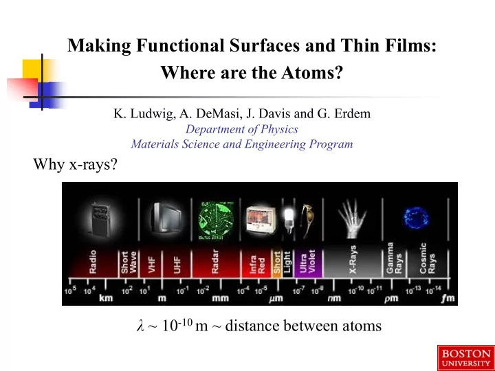
Department of Physics Materials Science and Engineering Program
Why x-rays? λ ~ 10-10 m ~ distance between atoms
SLIDE 2 X-ray Scattering (Diffraction)
The momentum transfer to an elastically scattered photon is:
images-mediawiki- sites.thefullwiki.org
2θ
SLIDE 3
X-ray Scattering (Diffraction)
In the Born Approximation, the intensity of x-ray scattering is equal to the structure factor S(q) of atom positions:
SLIDE 4
Thanks to synchrotron x-ray sources, available x-ray brightness has been growing fast – faster than computer speeds!
SLIDE 5
National Synchrotron Light Source (NSLS) and NSLS-II Brookhaven National Laboratory (Long Island)
SLIDE 6
National Synchrotron Light Source (NSLS) and NSLS-II Brookhaven National Laboratory (Long Island)
SLIDE 7 … the chambers can then be rolled
- nto the base diffractometer
permanently installed at NSLS Experimental conditions can be
- ptimized in ultra-high vacuum
(UHV) chambers at the home laboratory …
NSLS insertion device beamline X21 back hutch
Facility for Real-Time Studies of Surface and Thin Film Growth Processes
SLIDE 8
Surface Modification and Characterization Instrumentation
System installed on base diffractometer with UHV chamber, surface modification and characterization equipment
SLIDE 9
Flight Path Detector Incoming X-rays Incoming X-rays Exit X-rays Sample Sample manipulator
Facility for Real-Time X-ray Studies of Thin Film and Surface Processes
Physical Electronics PHI ion gun ∼ 2 x 10¹² ions/cm2s National Synchrotron Light Source (NSLS) – X21 Brookhaven National Laboratory
SLIDE 10
Grazing-Incidence Small-Angle X-ray Scattering (GISAXS)
qy qx
αi αf X
qz
ω 2θ X-rays Z Y detector
SLIDE 11
Grazing-Incidence Small-Angle X-ray Scattering (GISAXS)
SLIDE 12 BU Research Program
- Spontaneous Nano-Patterning of Surfaces during Ion Bombardment
– Aziz group (Harvard)
- III-V Nitride Semiconductor Growth (lasers, detectors, solar cells)
– T. Moustakas (BU), O. Malis (Purdue)
- Solid Oxide Fuel Cell (SOFC) Cathodes
– S. Basu, S. Gopalan, U. Pal (BU)
- Fundamental Processes in Thin Film Growth
– Headrick group (UVM)
SLIDE 13 Off-Axis Bombardment – Ripples: Normal Incidence Bombardment– Smoothening: Dots: GaSb (100)
ion direction
Si(100) Si(100)
Facsko et al., Science 285, 1551 (1999)
Spontaneous Nanopatterning of Semiconductor Surfaces by Ion Bombardment
SLIDE 14 Off-Axis Bombardment – Ripples: Normal Incidence Bombardment– Smoothening: Dots: GaSb (100)
ion direction
Si(100) Si(100)
Facsko et al., Science 285, 1551 (1999)
Fundamental questions:
- What physical processes cause nanopattern formation?
- How can we control it?
Importance of understanding patterning during bombardment:
- Hyperthermal bombardment is a ubiquitous technological process (e.g. sputter
deposition, ion-assisted deposition, PLD, sputter cleaning, plasma etching)
- Potential route for inexpensive nanopatterning of surfaces
SLIDE 15 Formation of ripple structures on surfaces during ion bombardment reported as early as 1960: Key early observations:
- Semiconductor surfaces are amorphized by ion bombardment at RT
- Wavelengths can be much longer than penetration depths of ions
Ar+ θ
5 keV Xe+ on graphite: Habenicht et al. PRB 60, R2200 (1999).
- At low bombardment angles,
ripple wavevector parallel to projected direction of ion beam
- At high bombardment angles, ripple
wavevector perpendicular to projected direction of ion beam
SLIDE 16 Theoretical treatment of Sputter Erosion – 1973:
Model of sputter erosion process:
- Energy deposited by incident ion
assumed to be a Gaussian ellipsoid
- Sputter rate from a given point on
the surface assumed to be proportional to energy deposited there
Chan and Chason, JAP 101, 121301 (2007)
SLIDE 17 Theoretical treatment of Sputter Erosion – 1973:
Surface Instability to Ion Bombardment due to curvature-dependent sputter erosion rate
Chan and Chason, JAP 101, 121301 (2007)
SLIDE 18 Theoretical treatment of Sputter Erosion – 1973:
Surface Instability to Ion Bombardment due to curvature-dependent sputter erosion rate
Chan and Chason, JAP 101, 121301 (2007)
Key Questions Not Addressed by Sigmund Mechanism:
wavelength?
- Why determines ripple
- rientation?
SLIDE 19 These are potentially answered by the Bradley-Harper Model – primary paradigm for last 20 years: Calculate local surface height evolution from Sigmund model to first
- rder in (ion penetration depth/surface radius of curvature):
2 2 2 2
2 1 2 1 ) , ( y h S x h S x h v t t r h
y x
∂ ∂ + ∂ ∂ + ∂ ∂ Γ + − = ∂ ∂ r
Average sputter erosion rate
SLIDE 20 These are potentially answered by the Bradley-Harper Model – primary paradigm for last 20 years: Calculate local surface height evolution from Sigmund model to first
- rder in (ion penetration depth/surface radius of curvature):
2 2 2 2
2 1 2 1 ) , ( y h S x h S x h v t t r h
y x
∂ ∂ + ∂ ∂ + ∂ ∂ Γ + − = ∂ ∂ r
Average sputter erosion rate Slope-dependent erosion rate causes motion of ripples across surface
SLIDE 21 These are potentially answered by the Bradley-Harper Model – primary paradigm for last 20 years: Calculate local surface height evolution from Sigmund model to first
- rder in (ion penetration depth/surface radius of curvature):
2 2 2 2
2 1 2 1 ) , ( y h S x h S x h v t t r h
y x
∂ ∂ + ∂ ∂ + ∂ ∂ Γ + − = ∂ ∂ r
Average sputter erosion rate Slope-dependent erosion rate causes motion of ripples across surface Curvature- dependent erosion rate
SLIDE 22
h B y h S x h S t t r h
y x 4 2 2 2 2
2 1 2 1 2 1 ) , ( ∇ − ∂ ∂ + ∂ ∂ = ∂ ∂ r
Stability or instability of surface for a ripple of a given wavelength is determined by tradeoff between curvature- dependent erosion rate and surface smoothening by diffusion/viscous flow Bradley- Harper: Add relaxation at short length- scales due to diffusion or viscous flow
SLIDE 23
- Lateral mass redistribution (CV) effect smoothens at low incidence angles
- Magnitude proportional to cos(2θ)
h j j t h ∇ ∝ ⋅ −∇ = ∂ ∂ v v
∂h ∂t = α∇2h → q2
SLIDE 24 More General Linear Theory Formalism
) , , ( 2 1 2 1 2 1
4 2 2
t y x h B h S h S t h
y y x x
η + ∇ − ∂ + ∂ = ∂ ∂
Linear theory growth or decay of local surface height fluctuations: Curvature coefficients Sx and Sy can include both curvature-dependent Bradley- Harper erosion instability and stabilization due to lateral mass redistribution
Uncorrelated Noise:
) , , ( = t y x η ) ' ( ) ' ( ) ' ( ) ' , ' , ' ( ) , , ( t t y y x x n t y x t y x − − − = δ δ δ η η
Ensemble-averaged height-height structure factor evolution:
[ ]
t q R t q R
e q R n q I e t q h t q h t q I
) ( ) (
1 ) ( ) ( ) , ( * ) , ( ) , ( − − + = =
( )
4 2 2
) ( Bq q S q S q R
y y x x
+ + − =
Amplification Factor:
SLIDE 25 Linear Theory of Surface Stability/Instability: Amplification Factor
If R(q) > 0, surface is unstable:
( )
4 2 2
) ( Bq q S q S q R
y y x x
+ + − =
Amplification Factor:
I(q,t)
t
If R(q)< 0, surface is stable: I(q,t)
t
) ( ) ( q R n q S >
I(q,t)
t
) ( ) ( q R n q S <
SLIDE 26 At each wavenumber, fit I(qx,y,t) to determine Amplification Factor R(qx,y) GISAXS evolution during 2 hours of 1 keV Ar+ bombardment
1 keV Ar+ on Si: For each ion bombardment angle θ use real-time GISAXS to measure I(qx,y,t)
Madi, Anzenberg, Ludwig and Aziz, PRL 106, 066101 (2011) Anzenberg, Madi, Aziz and Ludwig, PRB 84, 214108 (2011)
SLIDE 27
Measured Amplification Factor R(qx,y) as a function of incident angle in x- and y-directions
Transition from smoothening to ripple formation at ion bombardment angle θ ~ 45°.
SLIDE 28 Measured Amplification Factor R(q) as a function of incident angle in x- and y-directions
Simultaneously fit all R(qx,y) to form: R(qx) = -(Sx(θ)qx
2 + Bqx 4)
R(qy) = -(Sy(θ)qy
2 + Bqy 4)
to determine curvature coefficients Sx,y(θ)
SLIDE 29
Angular Dependence of Curvature Coefficients Sx,y(θ)
Fits show dominance of mass redistribution – both for smoothing at low angles and for creating ripples. Consistent also with simulations. (Kalyanasundaram et al., APL 92, 131909 (2008); Norris et al., Nature Comm. 10.1038/ncomms1280 (2011))
SLIDE 30
Data show dominance of Lateral Mass Redistribution through most of angular range
Above 45° ion incidence angle, roughening caused by ion momentum knocking surface atoms uphill !
Ion
Recoils Ions also knock atoms downhill when hitting downhill slope, but effective ion flux density is lower because of the slope…
SLIDE 31
X-ray Free-Electron Laser (XFEL)
In these studies, structure evolution has been on seconds time scale. Can we use x-rays to look at much faster time scales (e.g. motion of atoms in liquids)?
Self Amplified Spontaneous Emission (SASE) Femtosecond (10-15 s) x-ray laser pulses!
SLIDE 32
SLIDE 33
X-ray Free-Electron Laser (XFEL)
SLIDE 34
SLIDE 35 transversely coherent X-ray beam sample
X-ray Photon Correlation Spectroscopy (XPCS)
Study evolution of x-ray scattering “speckle” pattern to learn about motion of atoms, molecules
t1 t2 t3
monochromator “movie” of speckle recorded by CCD
g2(Δt) ≡ I(t) I(t + Δt) I
2
1
Δt g2 τ τ
−1(Q) = Rate(Q)
I(Q,t)
SLIDE 36
transversely coherent X-ray pulse from FEL sample
XPCS at LCLS using “Split Pulse” Mode
Femtoseconds to nanoseconds time resolution Uses high peak brilliance sum of speckle patterns from prompt and delayed pulses recorded on CCD
I(Q,Δt)
splitter variable delay Δt
Δt τ
Contrast
Analyze contrast as f(delay time)
10 ps ⇔ 3mm
SLIDE 37 transversely coherent X-ray beam sample
XPCS of Non-Equilibrium Dynamics using ‘Pumped’ Mode
- Femtoseconds to seconds time resolution
- Uses high peak brilliance
before Δt after pump
monochromator Correlate a speckle pattern from before pump to one at some Δt after pump Pump sample e.g. with laser, electric, magnetic pulse
SLIDE 38
Key Problem:
High peak x-ray beam intensity can damage sample (e.g. vaporize it!)
Damage to Ni-Pd-P samples by single pulse of LCLS x-ray beam
SLIDE 39
First Experiments Show Success is Possible
X-ray scattering from liquid Gallium
SLIDE 40 Conclusions
- Goal of condensed matter physics research is to understand
how materials behave and how new materials or material structures can be developed to meet societal needs
- The behavior of materials depends on the atomic structure and
evolution
- X-ray scattering is a uniquely powerful tool for determining
atomic structure (both static and evolving)
- Available x-ray intensities have been increasing dramatically
due to development of new sources (synchrotrons, FEL’s)
- X-ray scattering can give crucial information enabling us to
understand and control materials behavior