Lots of Issues What does increased breast density mean? Who - - PowerPoint PPT Presentation

lots of issues
SMART_READER_LITE
LIVE PREVIEW

Lots of Issues What does increased breast density mean? Who - - PowerPoint PPT Presentation

10/19/2018 Prevention and Screening for Breast Cancer: Whats New? Disclosures- I am a Medical Advisor and on the Scientific Advisory Board of the Mindy Goldman, MD company, Madorra, that is developing a Clinical Professor medical device


slide-1
SLIDE 1

10/19/2018 1

Prevention and Screening for Breast Cancer: What’s New?

Mindy Goldman, MD Clinical Professor

  • Dept. of OB/Gyn and Reproductive Sciences

Director, Gyn Center for Cancer Survivors and At-Risk Women UCSF Helen Diller Comprehensive Cancer Center and UCSF Women’s Health University of California, San Francisco

Disclosures- I am a Medical Advisor and on the Scientific Advisory Board of the company, Madorra, that is developing a medical device to treat vaginal dryness

Lots of Issues

Who should get MRI screening? What does increased breast density mean?

slide-2
SLIDE 2

10/19/2018 2

Breast Cancer Epidemiology- Issues pertaining to Screening

  • Approximately 12.3 % of women will be diagnosed with breast

cancer at some point during their lifetime. 1/8 at age 85

  • Number of new cases of breast cancer is 124.6 per 100,000

women per year

  • 14% all new cancers are breast
  • 12% diagnosed in reproductive years (ages 20-44)
  • More than 25,000 cases per year in California

Breast Cancer Epidemiology- Issues pertaining to Screening

  • Globally breast cancer is most common

diagnosed cancer in women and most common cause of cancer death

  • Almost 50% of breast cancers may be related

to risk factors

Breast Cancer- What do Clinicians Need to Understand?

  • What are the risk factors for breast cancer?
  • Who should be screened?
  • What type of screening should be done?
  • Who should be offered preventative medications or

surgeries?

Breast Cancer – Risk Factors

  • Age – increases as we age
  • Female gender
  • Race and ethnicity – highest incidence in whites, but

highest mortality in African Americans

  • Benign breast disease – particularly Atypical Ductal

Hyperplasia (ADH), Atypical Lobular Hyperplasia (ALH), or proliferative breast lesion. With atypia RR 3.7 to 5.3

slide-3
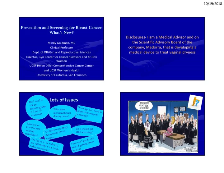
SLIDE 3

10/19/2018 3

Breast Cancer – Risk Factors

  • Family history and genetic factors – 15-20% of

women with breast cancer reported to have family history in first degree relative

  • Personal history of breast cancer increases

risk of contralateral breast cancer

  • Exposure to ionizing radiation

Breast Cancer – Risk Factors

  • Lifestyle and dietary factors

– May increase risk: obesity, smoking, high fat intake, red meat, alcohol use, soy phytoestrogens – May be protective: regular exercise, greatest benefit seen in adolescence – ?Vitamin D – some studies suggest low levels of Vit D associated with increased risk

Why is alcohol use associated with breast cancer?

  • Increases risk primarily of hormone positive cancer
  • Alcohol can affect the way estrogen is metabolized and increase

blood levels

  • Alcohol can reduce blood levels of folic acid which is involved in

copying and repairing DNA. Low levels of folic acid may make it more likely that DNA is incorrectly copied when cells divide causing errors that may lead to cancer

  • Women who have 2 - 5 drinks per day have about 1.5 x the risk of

developing breast cancer compared to non-drinkers

Breast Cancer – Risk Factors

  • Reproductive and hormonal factors

– Increased risk: early menarche, late menopause, late age of first child or nulliparity, increased breast density, long-term HRT, ?endogenous hormone levels – No association: prior abortion – Decreased risk: breastfeeding, ?Estrogen Replacement Therapy (ERT)

slide-4
SLIDE 4

10/19/2018 4

Breast Cancer – Risk Factors

  • Factors that may increase risk:

– Environment exposures : PCB’s, hair dyes, etc. - no clear data – Night time light exposure - Exposure to light at night suppresses nocturnal production of melatonin (thought to have antioxidant effects preventing damage to cells)

  • Factors without association:

– Antibiotic use – Caffeine – Antiperspirants – Bras – Breast implants

Breast Cancer – Risk Factors

  • Factors thought to be protective:

– Non-steroidal drugs - NSAIDs-especially aspirin and COX-2 inhibitors shown in recent meta- analysis to be protective for hormone positive breast cancer (likely via anti-inflammatory effects)

– de Pedro M, et al. Effect of COX-2 inhibitors and other non-steroidal inflammatory drugs

  • n breast cancer risk: a meta-analysis. Breast Cancer Res Treat. 2015 Jan;149(2):525-36

Breast Cancer Risk Factors

  • Bisphosphonates: meta-analysis have shown these

drugs can reduce the rate of breast cancer recurrence in bone and improve survival in postmenopausal women with breast cancer - not in premenopausal women and No clear data that it reduces the risk of getting breast cancer

Summarizing Risk

  • Most women can be categorized based on history
  • Risk prediction models can be used to stratify risk

if needed and take into consideration:

– Personal history of breast (and/or

  • varian/tubal/peritoneal ca)

– Family history of breast cancer – Known genetic mutation (like BRCA) – Breast density – Prior radiation to the chest was between ages 10-30

slide-5
SLIDE 5

10/19/2018 5

Issues pertaining to Screening?

  • How should screening should be done?
  • Should all women be screened?
  • What age to begin screening?
  • Should screening stop at older ages?
  • Should screening differ based on risk factors?

How Should Screening be Done?

  • Self Breast Exam (SBE)

– American Cancer Society no longer recommends – ACOG recommends teaching breast self awareness – Many other organizations (USPSTF) do not recommend – WHO and NCCN talk about SBE to “raise breast awareness” – Not great evidence that it helps

  • Few RCT, one of largest studies 2008 looked at 400,000

women in Russia and China showed no benefit in breast ca mortality and more biopsies done for benign disease

  • 2 case control studies showed less advanced disease if

women taught appropriate SBE

How Should Screening be Done?

  • Clinical Breast Exam (CBE)

– Many US organizations recommend CBE every 3 years from age 20 to 39, annually thereafter – American Cancer Society new guidelines do not support CBE – US Preventive Task force and WHO says not enough evidence to support regular CBE – ACOG says CBE can be offered with shared decision making – NCCN says annually for women over 40 – Evidence not clear:

  • most studies combine CBE with mammography so

independent effects less clear (only RCT did not show benefit)

  • Less standardization for CBE compared to mammography
  • 2009 review found no clear benefit

How Should Screening be Done?

  • Mammograms
  • Screening mammogram clearly detects early stage breast ca
  • Controversies are whether it improves breast cancer mortality (over

diagnosis) – 9 RCT looking at 650,000 women have looked at mortality – mammo +/- CBE. Results show a benefit in women ages 40-69 – Older studies looking at mortality also affected by older treatments that weren’t as effective – 25 yr. fu from randomized Canadian National Breast Screening Study did not show a benefit in mortality compared to exam alone when adjuvant therapy is available 22% (106/484) of screen detected invasive breast cancers were over-diagnosed

  • BMJ. 2014;348
slide-6
SLIDE 6

10/19/2018 6

Mammogram Screening Guidelines for Average Risk Women

  • All now talk about shared decision making
  • ACOG - every 1-2 years beginning at age 40
  • USPSTF -every 2 year screening should beginning at age 50
  • WHO - every 2 years for women 50-69
  • ACS -annually from 45-55 and every 2 years for women 55 and
  • lder
  • NCCN- annually at 40 with consideration of tomosynthesis

Why the Difference of Screening Mammo every 1-2 years?

  • Many North American groups recommend annual screening for

women under 55 because of evidence of more rapid tumor growth in younger women

  • Benefits of detecting more tumors in earlier stage in younger

women needs to be balanced against increased harms associated with an increased rate of false-positives

  • Every 2 year screening thought to decrease false positives, and

no overall differences in survival on a population basis

Mammogram Screening Common Areas

  • All groups state mammogram screening useful for women over

50

  • Groups agree that the benefits of mammography increase with

age

  • Once mammogram screening begins offer every 1-2 years
  • Differences in recommendations highlights need for

INDIVIDUALIZATION depending on risk

Should Mammogram Screening Stop at Older Ages?

  • Controversy as many cancers in women over 80 are slow

growing and not likely to affect survival, as other competing causes of death

  • 25% of breast cancer are diagnosed in women 75 and
  • lder but limited data on screening mammography in

this population

  • Groups now say that clinical judgment and predictive

models that combine age, comorbidities, and functional status can be used to identify women who may continue to benefit from screening mammography

slide-7
SLIDE 7

10/19/2018 7

Should Mammogram Screening Stop at Older Ages?

  • Differing guidelines:

– USPSTF states no benefit for screening above age 74 – ACR says continue until life expectancy < 5-7 years on basis of age or other co-morbidities – ACS continue screening mammography as long as their overall health is good and they have a life expectancy of 10 years or longer – ACOG,NCCN make no recommendations

  • CLINICIANS SHOULD INDIVIDUALIZE

Breast Screening with Newer Technologies

Need for data and subsequent guidelines

?

?

Other Screening Modalities - Breast MRI Breast MRI for Screening

  • Creates images of the breast by measuring changes in the

movement of protons in fat and water with changing magnetic fields. Image is created based on differences in tissue relaxation that occur after pulses of energy applied

  • Requires IV contrast with gadolinium and use based on

increased blood supply in tumors that take up and release of gadolinium quickly leading to specific pattern of rapid enhancement and washout on MRI

slide-8
SLIDE 8

10/19/2018 8

Breast MRI of invasive ductal cancer kinetic color map

2014 UpToDate

Breast MRI for Screening

  • Very sensitive for detecting small invasive cancers in high

risk women (but less specific than mammogram) with suggestion of improved mortality

  • No data showing a benefit for screening MRI in average risk

women

  • High false positives (nationally as high as 40%) which cause

patient anxiety, painful procedures

  • Substantial costs - $1000 (vs $100 for mammo)

Clinical Uses of Breast MRI

  • Assessment of silicone implant integrity
  • Follow up for occult breast cancers (missed on

mammogram) or assessing for primary breast ca in women with axillary disease

  • Assessing disease extent in newly diagnosed breast

cancer patients

  • Assessing response to neoadjuvant chemotherapy
  • Assessing recurrence of disease
  • Clarifying inconclusive clinical or mammographic

findings

  • Screening of high risk patients

MRI for Screening - Controversies

  • MRI can detect smaller cancers and more

node-negative malignancies in high risk women compared to other imaging modalities

  • No clear evidence of reduced mortality or

improved disease-free survival from screening with MRI

slide-9
SLIDE 9

10/19/2018 9

MRI Screening Recommendations in High Risk Women – ACS, NCCN

  • Annual MRI should be done in the following high-risk groups:

– Women with a BRCA1 or BRCA2 mutation – Women who have a first-degree relative with a BRCA mutation (even if they have not yet been tested themselves) – Prior radiation therapy to the chest between the ages of 10- 30 – Known Li-Fraumeni syndrome, Cowden syndrome, or Bannayan-Riley-Ruvalcaba syndrome, or have first-degree relatives with one of these syndromes – Women with an approximate lifetime risk of breast cancer from 20 -25%, according to risk prediction models primarily using family history

MRI for Screening

  • No clear benefits of MRI screening in low or

average risk women

  • The American Cancer Society and other

groups recommend against MRI screening for women whose lifetime risk of breast cancer is less than 15%

  • Weight gain during this time of year is felt to be

due to…..

  • 1. Eating more (duh) in the fall
  • 2. Lack of sunshine
  • 3. Being more of a “coach potato” when it starts

to get cold

And the Correct Answer is…..

  • Lack of sunshine - Researchers have found

that lack of vitamin D reduces fat breakdown and can trigger fat storage. So, the lack of sunlight has more to do with the extra bit around the waist or hips than all the pumpkin spice lattes. Well, at least some of it …….

slide-10
SLIDE 10

10/19/2018 10

Other Screening Modalities

Tomosynthesis “3D” Mammography

  • Modification of digital mammography that uses a moving x-

ray source and digital detector to create 3-D view

  • Currently FDA approved for clinical use as an adjunct to

mammography in most places, but on April 9, 2018, FDA accredited digital breast tomosyntheis systems in Arkansas, Iowa ,Texas, and the American College of Radiology (ACR)

  • Thought to decrease recall rates by delineating true lesions

from superimposition

Tomosynthesis “3D” Mammography

  • RCT, prospective and several retrospective studies comparing

digital mammo alone vs digital mammo plus breast tomosynthesis found improved positive predictive value for cancer, lower recall rates

  • incremental breast cancer detection is in the range of 0.5-

2.7/1000 screens

  • Recall rates in the range of 0.8-3.6%.

– Radiology. 2018 Jun;287(3):787-794. – Eur Radiol. 2018 Aug 29. – Lancet Oncol. 2016 Jun 23

Tomosynthesis “3D” Mammography

  • Older techniques had twice the radiation exposure
  • Newer techniques create a 2-D image from the 3-D tomo

images, lowering radiation dose to slightly above or equal standard mammogram

  • Particularly useful for moderately dense breasts and high risk

women

  • Need more RCT’s before recommending for all women
slide-11
SLIDE 11

10/19/2018 11

Whole Breast Ultrasound

  • Useful for dense breast tissue, but increased false positives
  • Ultrasound in addition to mammography slightly improves

cancer detection but has not been shown to affect breast cancer mortality

  • No RCT comparing screening ultrasound plus mammo vs.

mammo alone

  • Operator dependent

Whole Breast Ultrasound

  • Confusion for clinicians and patients as many patients have

been screened in centers where ultrasound is routinely done, but no guidelines for use in screening

  • FDA approved an automated device in 2012 for as an adjunct to

mammography for asymptomatic women with dense breasts and a negative mammogram – no published data from a screening trial using this device

  • Generally felt not enough evidence to support device

Molecular breast imaging (MBI)

  • Uses small, semiconductor-based gamma cameras to image the

breast following injection of a radiotracer. Cancers absorb this tracer and “light up”

  • Other names for technique: Miraluma test, sestamibi,

scintimammography, or breast specific gamma imaging

  • FDA approved 1999 and is used as an adjunct to mammography

to help differentiate scar tissue from cancer recurrence in patients with prior diagnosis, to screen high-risk patients who are contraindicated for MRI and to help patients with indeterminate findings on mammography

Molecular breast imaging (MBI)

  • Studies at Mayo Clinic study in 2008 and 2015 showed much

more sensitivity for cancer detection compared to mammo alone (most recent study 360% increased detection rate)

  • Particularly useful for women with dense breasts
  • Much higher dose of radiation - half the average U.S. yearly

exposure

  • Ongoing trials comparing MBI to MRI
  • Not recommended in low-average risk women
slide-12
SLIDE 12

10/19/2018 12

Computer Aided Detection (CAD)

  • Computer-based technology designed to recognize

mammographic patterns and help radiologists identify suspicious areas

  • FDA approved CAD in 1998 after several studies showed

improved cancer detection

  • Debate about usefulness and no randomized trials have been

performed to determine its effect on breast cancer mortality

  • Picks up more DCIS
  • Studies have shown improved sensitivity but higher recall rate

and potential for over-diagnosis

Thermography

  • Developed based on the observation that patients have

elevated breast skin temperatures over their breast cancers

  • Received FDA approval in 2004 based on safety of infrared

imaging technology but not based on efficacy

  • Many thermography centers now exist
  • Specificity is low
  • No studies have shown this to be an effective screening tool

Thermography

  • No major organization makes screening

recommendations for thermography

  • FDA issued a safety communication in June 2011

saying thermography is not a replacement for screening mammography and on its own is not an effective screening tool - difficult when abnormal thermogram and normal mammogram

Breast Screening – my clinical pearls for clinicians

  • Not enough evidence to support teaching SBE
  • Evidence is mixed whether to do CBE at routine visits
  • Baseline mammograms 40-50 depending on risks
  • Frequency of mammography 1-2 years
  • Continue mammogram screening as long as patient

is healthy

  • Do not use MRI screening in average risk women
  • Not enough evidence for adjuvant use of newer

imaging techniques

  • INDIVIDUALIZE recommendations based on risk
slide-13
SLIDE 13

10/19/2018 13

More Fun Facts!

  • Fall colors are caused by…..
  • 1. Hallucinations as more people do drugs in

the fall compared to other seasons

  • 2 Red pigment in leaves
  • 3. Sugar in leaves

And the Correct Answer is…..

The more red in the leaf, the more sugar the leaf is storing. That is why Maple trees are so vibrant. Evergreens don’t change because their leaves have a thick wax covering that protects the green chlorophyl in the leaves.

Prevention of Breast Cancer: Clinical Issues

  • When should we consider?

– Drugs: Tamoxifen, Raloxifene, Aromatase Inhibitors – Preventative Surgeries: Prophylactic mastectomies, oophorectomies

Prevention of Breast Cancer: Clinical Issues

  • Consider based on risk- ASCO and USPSTF have guidelines

– High risk based on family history: particularly first degree relatives diagnosed pre-menopausal or multiple family members – age > 35 – Risk Prediction Models like Gail, BCSC: > 1.7% 5 yr. risk – Concern for hereditary breast ovarian cancer syndrome – A history of lobular carcinoma in situ (LCIS) or prior biopsies showing atypical change

slide-14
SLIDE 14

10/19/2018 14

Tamoxifen for Breast Cancer Prevention

  • Complex drug with estrogen and anti-estrogen properties
  • Approved by FDA in 1998 for breast cancer prevention in high risk

pre and PMP women

  • 2013 US Preventive Services Task Force (USPSTF) meta-analysis of

4 trials showed 30% reduction in risk of primarily hormone positive breast ca in tamoxifen users vs. placebo (RR 0.70, 95% CI 0.59-0.82)

  • Treatment trials have shown a greater benefit for 10 yrs. vs 5 yrs.
  • f use but no data in prevention setting so recommendation to

take for 5 years

Raloxifene for Breast Cancer Prevention

  • In the same class of drugs as Tamoxifen
  • Raloxifene - FDA approved 2007 for prevention of breast cancer

in PMP women

  • 2013 US Preventive Services Task Force (USPSTF) meta-analysis
  • f 4 trials showed 10% reduction in risk of hormone positive

breast ca in raloxifene users vs. placebo (nine cases in 1000 women, RR 0.90, 95% CI 0.27-0.71)

  • No increased incidence of endometrial cancer
  • No difference in breast cancer-specific or all-cause mortality

Tamoxifen and Raloxifene - overall data

  • Overall in 7 fair/good quality studies, tamoxifen and

raloxifene reduced incidence of invasive breast cancer by 7 to 9 cases in 1000 women over 5 years compared with placebo- in 1 study tamoxifen slightly more effective than raloxifene

  • Neither tamoxifen or raloxifene trials showed difference in

breast cancer-specific or all cause mortality

  • Fewer thromboembolic events, cataracts, and endometrial

cancer with ralxoifene

Aromatase Inhibitors in the Prevention Setting

  • Tamoxifen competes with estrogen at receptor binding site in

the breast preventing receptor activation

  • Aromatase Inhibitors prevent conversion of male hormones

into estrogens and decrease peripheral circulating estrogen

  • Third generation AI’s used:
  • Anastrozole (Arimidex)
  • Letrozole (Femara)
  • Exemestane(Aromasin)
slide-15
SLIDE 15

10/19/2018 15

Aromatase Inhibitors in the Prevention Setting

  • International Breast cancer Intervention Study (IBIS-II), looked

at 4000 PMP women at high risk of breast cancer and randomly assigned them to treatment with anastrozole vs. placebo for 5 yrs. Found 50% percent reduction in the number of invasive breast cancers or DCIS with AI vs. placebo [HR] 0.47, 95% CI 0.32-0.68

  • American Society of Clinical Oncology (ASCO) added this to

updated guidelines in 2013 for prevention of hormone positive breast cancer in PMP women

  • Not yet FDA approved in the prevention setting

Preventative Surgeries for Reducing Risk of Breast Cancer

  • Risk Reducing Mastectomies (RRM)

– Removes most, but not all of breast tissue

  • Skin Sparing procedures

– Recently developed procedure – Removes more breast tissue than subcutaneous mastectomy – Usually combined with immediate reconstruction – Provides good cosmetic result

Do Preventative Surgeries Prevent Breast Cancer?

  • Significantly reduces risk of breast cancer and death
  • In premenopausal women, prophylactic mastectomies

decreases the risk of hormone receptor positive breast cancer by 90+%

  • For women having prophylactic oophorectomy before age 40,

there is an approximate 50% reduction in breast cancer risk

  • Recommendation to consider in BRCA or other gene mutation

carriers that carry significant increased lifetime risks for breast cancer

Clinical Concerns

slide-16
SLIDE 16

10/19/2018 16

Should mammograms still be done if women get MRI for screening? YES

  • Mammograms can pick up calcifications not

seen on MRI that could be a sign of cancer

  • High risk women should begin MRI screening

annually starting at 25 and then alternate annual MRI and mammogram starting at 30

Is breast imaging needed after bilateral mastectomies? NO

  • If complete mastectomy no need for imaging
  • If skin sparing, nipple sparing procedure, some centers may still

recommend screening mammography

  • MRI can be helpful to establish the presence of residual breast

tissue after bilateral mastectomy, and routine screening not recommended if no residual breast tissue is seen

  • With saline or silicone implants or autologous reconstruction

procedures imaging typically not recommended

Conclusions

  • Hopefully you now will now:

– Know the risk factors for breast cancer – Understand issues and controversies surrounding screening from a clinicians perspective – Know what types of breast screening exist – Know what drugs are used for prevention – Know what the screening and prevention strategies are for women who are high risk – Understand that screening and prevention should be individualized