SLIDE 1
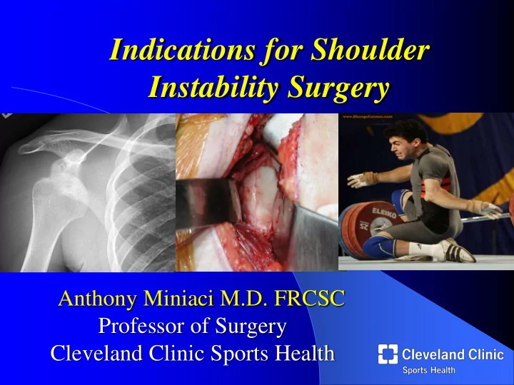
Indications for Shoulder Instability Surgery
Anthony Miniaci M.D. FRCSC Professor of Surgery Cleveland Clinic Sports Health
SLIDE 2 Considerations
- Instability classification
- Natural history
- Patient demographics-age,
activity level, occupation, associated morbidities
- Associated pathology
- Prior treatments
Indications for Shoulder Instability Surgery
SLIDE 3 Instability Classification: Spectrum
- Unidirectional
- “Classic” MDI
- More surgery on “MDI”
- Multidirectional instability
lacks definitions-”MDI” has grown?
SLIDE 4
Shoulder Instability
Surgery…………………………..Rehabilitation
SLIDE 5 Multidirectional Instability
Lack of definitions
- 1. Patient population
- 2. Symptom
definition
SLIDE 6 Multidirectional Instability
Patient Population
voluntary, involuntary, habitual
posterior,multi
trauma, atraumatic (“born, torn, worn loose”)
- Laxity- degree, focal vs.
general, symmetry
Voluntary, positional, Posterior,Traumatic Multi, Involuntary, Atraumatic Anterior, Involuntary Atraumatic
SLIDE 7
- 2. Symptom Definition
- pain, Instability or Both
- Instability- degree
(subluxation/dislocation)
microinstability
Multidirectional Instability ! Laxity vs. Instability
source of Sx
AC joint, etc.
with subluxation
SLIDE 8
The MDI patient pathology- Imaging Studies
Labral Tears Rotator cuff tears Capsular laxity Leakage of dye
SLIDE 9
- Best results in milder forms of
instability (subluxation), multidirectional laxity, anteroinferior instability with MD laxity
- Good for patients with PAIN and
laxity
- Associated pathology best treated
arthroscopically
Arthroscopic Capsular Shifts
SLIDE 10 What about Posterior Instability?
- Dislocation
- Fixed, Chronic
- Recurrent
- Subluxation
- Traumatic
- Micro
- macro
- Muscular- Voluntary
- Scapula winging
- Asymmetric muscular contraction
- Positional- Involuntary
SLIDE 11 What about Posterior Instability?
- Dislocation
- Fixed, Chronic
- Recurrent
- Subluxation
- Traumatic
- Micro
- macro
- Muscular- Voluntary
- Scapula winging
- Asymmetric muscular contraction
- Positional- Involuntary
SLIDE 12 What about Posterior Instability?
- Dislocation
- Fixed, Chronic
- Recurrent
- Subluxation
- Traumatic
- Micro
- macro
- Muscular- Voluntary
- Scapula winging
- Asymmetric muscular contraction
- Positional- Involuntary
SLIDE 13
What about Posterior Dislocation ?
SLIDE 14
Posterior Dislocation- Arthroscopy
Reverse Hill Sachs Lesion Posterior Glenoid
SLIDE 15
SLIDE 16 Posterior Instability/Subluxation
- Repetitive microtrauma → posterior
labral/ligamentous/capsular attenuation & tearing
RPS – Recurrent Posterior Subluxation
- Weightlifters, Linemen, tennis players,
throwers, swimmer
- Present with vague and diffuse posterior
shoulder pain
- Not typical to have recurrent dislocations
- Pathoanatomy
- posterior labrum, post capsule and
posterior part of IGHL
SLIDE 17 Treatment
Non operative
- Rehabilitation, biofeedback,
proprioceptive training
- Cuff, deltoid, periscapular
strengthening
Surgical
arthroscopic/open capsulolabral reconstruction
- Arthroscopic labral repair
- Anatomic, minimally invasive,
effective
SLIDE 18 Anterior Instability Epidemiology
- Most common
- Bimodal distribution
- 2nd & 6th decades
- 96% result from traumatic event
- Overall incidence 1.7%
- ~3 times incidence in males
Rowe et al., ‘56; Hovelius et
- al. ‘82
- Recurrent Shoulder Instability
- Age-related recurrence following traumatic anterior instability
- < 20 years old – 66-94%
- 20 to 40 years old – 40-74%
- > 40 years old – 10%- ROTATOR CUFF tears
SLIDE 19 Recurrent Instability
Associated pathology
Osteoarthritis
- Hovelius JSES ’09
- Nonoperative tx with 1 more recurrence had
more arthropathy
- Buscayret AJSM ‘04
- number of dislocations linked to risk for
postop OA
- Ogawa AJSM ‘10
- 5-20 yr f/u study of 167 open Bankarts
evaluated with pre and postop CT
- Postop OA correlates with increased number
- f instability events
SLIDE 20 Arthroscopy
- 127 patients; diagnostic arthroscopy, x-rays (AP, Glenoid,
West Point), MRI
SLIDE 21 Anterior Shoulder Instability
Surgical Pathology
- Labral tears
- Capsular redundancy
Unrecognized Pathology
Deficiencies/Avulsions
- Labral pathology/ALPSA
- Subscapularis tears- Open
- BONE LOSS
- Glenoid erosion or
fractures(Bankart)
SLIDE 22 Treatment options
Glenoid Bone Loss
- 1. Bristow /Latarjet
- 2. Bone Graft- auto/allo
Humeral Bone Loss
- 1. Remplissage
- 2. Decreased ER
- 3. Arthroplasty
SLIDE 23 Glenoid Bone Loss
arthroscopically/ soft tissue/open
- Larger defect requires
- pen fixation if bone
stock adequate
- Quantify with 3D CT
- Consider combined
effect of a humeral defect- treat smaller lesions
Provencher et. al. JBJS, 2010
Bois, Jones, Miniaci et. al. AJSM, 2012
SLIDE 24 Glenoid Bone Loss
arthroscopically/ soft tissue/open
- Larger defect requires
- pen fixation if bone
stock adequate
- Quantify with 3D CT
- Consider combined
effect of a humeral defect- treat smaller lesions
Provencher et. al. JBJS, 2010
Bois, Jones, Miniaci et. al. AJSM, 2012
SLIDE 25
What about the Hill Sachs ?
SLIDE 26 Hill Sachs
must be addressed
- Remplissage
- French for “to fill”
- Infraspinatus tucked into
defect
when defect is >25%
Provencher et al., JAAOS, 2012
SLIDE 27 Hill Sachs
must be addressed
- Remplissage
- French for “to fill”
- Infraspinatus tucked into
defect
when defect is >25%
Provencher et al., JAAOS, 2012
SLIDE 28 2013 Anatomic Reconstruction Options
- Matched Allograft
- Autogenous Iliac crest
Autograft
Option
reported 0% redislocation
SLIDE 29
Combined Humeral Head and Glenoid Reconstruction
SLIDE 30 Shoulder Instability Surgery Summary
- Know your patient
- Demographics, activity
etc
- Know the pathology
- Type of instability
- Associated pathology
- Know the natural
history
SLIDE 31
Anthony Miniaci M.D. FRCSC Professor of Surgery Cleveland Clinic