
SLIDE 1
1
Approach to the Adnexal Mass
UCSF Helen Diller Cancer Center
Approach to the Adnexal Mass
Stefanie M. Ueda, M.D.
Assistant Clinical Professor Division of Gynecologic Oncology University of California, San Francisco
Approach to the Adnexal Mass
UCSF Helen Diller Cancer Center
I have nothing to disclose.
Approach to the Adnexal Mass
UCSF Helen Diller Cancer Center
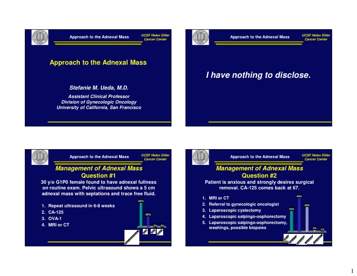
Management of Adnexal Mass Question #1
30 y/o G1P0 female found to have adnexal fullness
- n routine exam. Pelvic ultrasound shows a 5 cm
adnexal mass with septations and trace free fluid.
64% 3% 3% 30%
- 1. Repeat ultrasound in 6-8 weeks
- 2. CA-125
- 3. OVA-1
- 4. MRI or CT
Approach to the Adnexal Mass
UCSF Helen Diller Cancer Center
Management of Adnexal Mass Question #2
Patient is anxious and strongly desires surgical
- removal. CA-125 comes back at 67.
25% 40% 1% 3% 30%
- 1. MRI or CT
- 2. Referral to gynecologic oncologist
- 3. Laparoscopic cystectomy
- 4. Laparoscopic salpingo-oophorectomy
- 5. Laparoscopic salpingo-oophorectomy,