Hand-held dual-particle imager development based on Stilbene array - - PDF document

hand held dual particle imager development based on
SMART_READER_LITE
LIVE PREVIEW

Hand-held dual-particle imager development based on Stilbene array - - PDF document

Transactions of the Korean Nuclear Society Virtual Spring Meeting July 9-10, 2020 Hand-held dual-particle imager development based on Stilbene array coupled with SiPM array Jihwan Boo, Seoryeong Park and Manhee Jeong Nuclear & Energy


slide-1
SLIDE 1

Hand-held dual-particle imager development based on Stilbene array coupled with SiPM array

Jihwan Boo, Seoryeong Park and Manhee Jeong Nuclear & Energy Engineering Dept., Jeju Nat. Univ., 102 Jejudaehak-ro, Jeju-si, Jeju-do, 63243

*Corresponding author: mhjeong@jejunu.ac.kr

  • 1. Introduction

Dual-particle imagers (DPI) using coded-aperture mask or scatter camera are widely used in the homeland security and nuclear decommissioning fields for the purpose of localization and determining of unknown radionuclide [1-2]. In particular, securing images of neutrons and gamma rays in the nuclear decommissioning process is important in that it makes it easier for the workers to classify the waste generated during the work and can reduce costs effectively. The DPI developed so far is mainly composed of imaging techniques that apply scatter camera, spatial coded-aperture, and time-encoded imaging methods using the liquid scintillator or organic scintillators such as plastic and stilbene. In these imaging devices, it is important to separate neutrons from gamma rays, and various methods have been studied. Neutrons and gamma rays can be separated by pulse shape discrimination (PSD) methods. Methods for distinguishing neutrons from gamma rays include the charge comparison method (CCM), the simplified charge comparison method (SCCM), the pulse gradient analysis (PGA), and the neutron gamma model analysis (NGMA) [3]. Of these methods, the charge-integration method is widely used because the method is simple and has superior performance compared to other methods. Although this method is excellent for discrimination of high energy gamma-rays and neutrons, it is difficult to distinguish between gamma-rays and neutrons for low energy (below ~500 keVee neutron energy deposed), making it inevitable for misclassification [4]. Nuclear imaging can be performed by combining a detector with a conventional real-image camera that can distinguish neutron and gamma-ray reactions using these PSD techniques. It provided images of radioactive hotspot locations, enabling them to identify the characteristics of the nuclear sites. However, there are some drawbacks to the technology currently used to perform on-site characterization. The identification process can take a long time, as commonly used detectors and imagers must complete a slow scan process before results can be obtained. Therefore, in this paper, we would like to describe the development of the Stilbene array based hand-held type dual-particle imager and the characteristics of its system, which allows us to overcome current shortcomings and easily separate events of gamma rays and neutrons simultaneously to acquire each image.

  • 2. Methods and Results

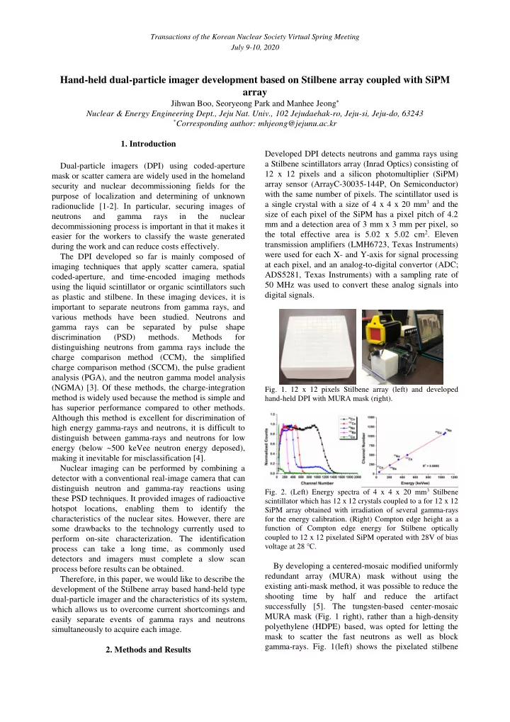
Developed DPI detects neutrons and gamma rays using a Stilbene scintillators array (Inrad Optics) consisting of 12 x 12 pixels and a silicon photomultiplier (SiPM) array sensor (ArrayC-30035-144P, On Semiconductor) with the same number of pixels. The scintillator used is a single crystal with a size of 4 x 4 x 20 mm3 and the size of each pixel of the SiPM has a pixel pitch of 4.2 mm and a detection area of 3 mm x 3 mm per pixel, so the total effective area is 5.02 x 5.02 cm2. Eleven transmission amplifiers (LMH6723, Texas Instruments) were used for each X- and Y-axis for signal processing at each pixel, and an analog-to-digital convertor (ADC; ADS5281, Texas Instruments) with a sampling rate of 50 MHz was used to convert these analog signals into digital signals.

  • Fig. 1. 12 x 12 pixels Stilbene array (left) and developed

hand-held DPI with MURA mask (right).

  • Fig. 2. (Left) Energy spectra of 4 x 4 x 20 mm3 Stilbene

scintillator which has 12 x 12 crystals coupled to a for 12 x 12 SiPM array obtained with irradiation of several gamma-rays for the energy calibration. (Right) Compton edge height as a function of Compton edge energy for Stilbene optically coupled to 12 x 12 pixelated SiPM operated with 28V of bias voltage at 28 ℃.

By developing a centered-mosaic modified uniformly redundant array (MURA) mask without using the existing anti-mask method, it was possible to reduce the shooting time by half and reduce the artifact successfully [5]. The tungsten-based center-mosaic MURA mask (Fig. 1 right), rather than a high-density polyethylene (HDPE) based, was opted for letting the mask to scatter the fast neutrons as well as block gamma-rays. Fig. 1(left) shows the pixelated stilbene

Transactions of the Korean Nuclear Society Virtual Spring Meeting July 9-10, 2020

slide-2
SLIDE 2

scintillators and their mechanical coupling to the data acquisition (DAQ) system. As shown in Fig. 2, the energy spectrum was acquired using various gamma-ray sources, and energy corrections to units of keVee are made in where the coefficient of determination (R2) for ADC values was 0.9995 in the range of Compton edges

  • f 43.485 keV to 1061.71 keV.
  • Fig. 3. 2D flood histogram of 12 x 12 pixels Stilbene

scintillator array for 60Co, 137Cs, 22Na, 133Ba, and 57Co gamma ray irradiation.

As shown in Fig. 3, all of the 144 detecting elements can be identified. However, when the gamma-ray energy decreases, positional resolution across the array was degraded because the Compton scattering angle was

  • larger. This increases the uncertainty of the interaction

localization, but it enhances detection efficiency, which can minimize the exposure time. With respect to decoding a detector pattern, this negative impact can be mitigated as far as most scattered events are taken in a pixel without pixel jumping.

  • Fig. 4. Illustration of the implementation of the pulse shape

discrimination method used in this study. Long and short integrals used in CCM calculations are clearly marked on the plot.

  • Fig. 4 shows pulses generated in response to the

neutron and γ-ray interaction in stilbene scintillators, where the gamma-ray pulses decay much faster than

  • neutron. For each detected waveform, the integrated

charge of the short and long gates was recorded and a PSD value was calculated: a ratio between one over the tail of the pulse (the tail integral) and one over the entire pulse (the total integral). The PSD scatter plot can then be obtained, as shown in Fig. 5, and a clear distinction between neutron and gamma-ray induced events are

  • represented. The gammas are found between PSD

values of around 0 to 0.2. Above this, in the second cluster, are the fast neutrons. None of the curvatures of each cluster at higher integrated charge values is

  • bserved employing the low-precision ADC, unlike
  • ther neutron detector systems based on stilbene crystal

coupled to photo-multiplier tube (PMT) [4]. It is postulated that linearity over amplification of the induced detector signal output from the SiPM resulted in this positive impact, by performing a linear gain correction for every pixel [5]. The partial overlap between the gamma and neutron clouds is recorded in the range below 200 keVee, suggesting an inevitable degree of misclassification of the events.

  • Fig. 5. PSD discrimination plots for 144 pixels using CCM

method (left) and PSD plot at full energy range (right)

  • btained with the CCM in the optimized set-up, using the

Stilbene scintillator measuring 252Cf and 137Cs sources.

  • Fig. 6. The main graphical user interface (GUI) view of

system operation which displays the position and count rate of the 252Cf spontaneous fission source at 50 cm distance from center with 30 seconds detection time.

Following each neutron and gamma-ray interaction, the system develops a signal from the SiPM array using a symmetric charge division (SCD) circuit, from which direct readout of 12 x 12 rows and columns is applied in

  • rder to determine the energy deposited and the

centroidal position of the interacting pixel. That information is then displayed on the image plane using MLEM image reconstruction techniques. This radiographic mapping is overlaid in real-time with an

  • ptical image derived from a complementary metal-
  • xide-semiconductor (CMOS) image sensor as shown in
  • Fig. 6.

Transactions of the Korean Nuclear Society Virtual Spring Meeting July 9-10, 2020

slide-3
SLIDE 3
  • 3. Conclusions

A combination of the photon and neutron imaging systems is particularly appealing to monitor the flow of materials of interest, such as undeclared activities involving special nuclear material. Coded-aperture based DPI can be totally beneficial when trying to detect a highly enriched uranium under active interrogation, using both fission neutrons and prompt photons in the presence of various types of shielding [1- 2]. In the coded-aperture based dual-particle imaging system will be used to detect the accurate location of unknown and shielded radiation sources in real-time and to develop equipment for nuclide analysis in the field of medical, nuclear industrial, and homeland security. Acknowledgements This work was partly supported by Korea Institute of Energy Technology Evaluation and Planning (KETEP) grant funded by the Korea government (MOTIE) (20181520302230) and by the Nuclear Safety Research Program through the Korea Foundation of Nuclear Safety (KoFONS) using the financial resource granted by the Nuclear Safety and Security Commission (NSSC)

  • f the Republic of Korea (No. 1903011-0119-CG100).

REFERENCES

[1] M. C. Hamel, J. K. Polack, M. L. Ruch, M. J. Marcath, S.

  • D. Clarke, M. Flaska, and S. A. Pozzi, Active neutron and

gamma-ray imaging of highly enriched uranium for treaty verification, Scientific reports, Vol. 7, pp. 1-10, 2017. [2] J. K. Polack, A. Poitrasson-Rivière, M. C. Hamel, K. Ide,

  • K. L. McMillan, S. D. Clarke, M. Flaska, and S. A. Pozzi,

Dual-particle imager for standoff detection of special nuclear material, IEEE Nuclear Science Symposium Conference Record, pp. 1494-1500, 2011. [3] P. A. Hausladen, M. A. Blackston, E. Brubaker, D. L. Chichester, P. Marleau, and R. J. Newby, Fast-neutron coded- aperture imaging of special nuclear material configurations, Report INL/CON-12-26506, Idaho National Laboratory (INL), 2012. [4] P. Kerr, and V. Mozin, Stilbene Scintillator Detector Array and Data-Acquisition System for Active Interrogation Report No. LLNL-TR-757673, Lawrence Livermore National Lab (LLNL), 2018. [5] M. Jeong, B. Van, B. Wells, L. D’Aries, and M. Hammig, Scalable Gamma-ray Camera for Wide-Area Search Based on Silicon Photomultipliers Array, Review

  • f

Scientific Instruments, Vol. 89, pp. 033106, 2018. Transactions of the Korean Nuclear Society Virtual Spring Meeting July 9-10, 2020