
SLIDE 1
4/21/2018 1
Protective Role of Fibroblast Growth Factor Signaling in Hypoxia-Induced Pulmonary Hypertension
Kel Vin Woo, M.D., Ph.D. Pediatric Cardiology, Fellow Ornitz Lab UCSF 11th International Conference Neonatal & Childhood Pulmonary Vascular Disease April 21st, 2018
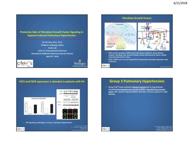
Fibroblast Growth Factors
Ornitz and Itoh.Dev. 2015
- FGF2 and its receptors FGFR1 and 2 have been studied in many disease
models, including: skin wound healing, retinal microvascular injury, cardiac ischemia/reperfusion injury.
- FGF2, FGFR1 and 2 are not required for homeostasis but play important roles
in disease.
Control PH
FGF2 and FGFR expression is elevated in patients with PH
1. Izziki et al. JCI 119. 2009 2. Tu et al. Am J Respir Cell Mol Biol 45. 2011 3. Kim et al. Nat Med 19(1). 2013 Pulmonary Hypertension Control
Lung tissue of PH patients
- FGF signaling is pathologic in Group 1 pulmonary hypertension.
Group 3 Pulmonary Hypertension
- Group 3 (2nd most common): Alveolar hypoxia due to lung diseases
including bronchopulmonary dysplasia (BPD), interstitial lung disease, COPD, OSA, alveolar hypoventilation disorders, chronic exposure to high altitude.
1.Osterman. Pediatrics 135(6). 2015. 2.Stoll. Pediatrics 126(3). 2010. 3.Mourani. AJRCCM (191). 2015.
- 4. Khemani. Pediatrics 120. 2007.