Glucocorticoid Associated Osteoporosis 1950 Raoul Dufy - - PowerPoint PPT Presentation

glucocorticoid associated osteoporosis
SMART_READER_LITE
LIVE PREVIEW

Glucocorticoid Associated Osteoporosis 1950 Raoul Dufy - - PowerPoint PPT Presentation

The Many Benefits of Glucocorticoids Glucocorticoid Associated Osteoporosis 1950 Raoul Dufy Prevention and Treatment Tomorrow I sail to Boston in search/Of my Jonathan Graf, MD hands. . . . Fifteen years ago the attacks began. Disfiguring


slide-1
SLIDE 1

1

Glucocorticoid Associated Osteoporosis

Prevention and Treatment

Jonathan Graf, MD Professor of Clinical Medicine UCSF Division of Rheumatology, San Francisco General Hospital

The Many Benefits of Glucocorticoids

Tomorrow I sail to Boston in search/Of my

  • hands. . . . Fifteen years ago the attacks
  • began. Disfiguring my

hands/As if I were painting with leaden

  • gloves. . . . My only wish: to draw

freehand/Following the wind/ Flowers that beckon me/And capturing them/In a vase of Anemones.

1950 Raoul Dufy 1951 ”La Cortisone”

And the drawbacks….

1952 1951-1953 Dufy finished over 200

  • paintings. But he passed away due

to a GI hemorrhage at least partially caused by his therapy
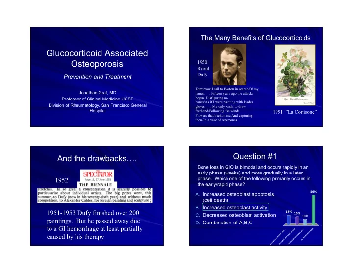
Question #1

Bone loss in GIO is bimodal and occurs rapidly in an early phase (weeks) and more gradually in a later

  • phase. Which one of the following primarily occurs in

the early/rapid phase?

  • A. Increased osteoblast apoptosis

(cell death)

  • B. Increased osteoclast activity
  • C. Decreased osteoblast activation
  • D. Combination of A,B,C

Increased osteoblast apo... Increased osteoclast activity Decreased osteoblast act... Combination of A,B,C

18% 56% 10% 15%

slide-2
SLIDE 2

2

Glucocorticoids: Some toxicities

Diabetes Cataracts HTN Weight gain Fluid retention PUD Myopathy Psychiatric

OSTEOPOROSIS & OSTEONECROSIS

50% of patients

HIGH MEDIUM CONTROL

Steroids

WOMEN

MEN Fracture risk increases with AGE, GENDER, and Dose

*

Van Staa et al, JBMR, 1998

Glucocorticoid Effects of Bone

Increased apoptosis of

  • steocytes (bone quality

decreased before BMD) Prolonged osteoclast survival and decreased

  • steoclastogenesis

Increased Osteoclast bone resporption Decreased

  • steoblastogenesis and

increased apoptosis

Multiple pathways affected!!!! Increased bone resportion relative to decreased bone formation and decreased overall bone quality:

Weinstein R. NEJM 2011;365:62-70

GLUCOCORTICOIDS: “Double Whammy to Bones”

Osteoclast /Pro-resorptive effects (early)

– Decrease OPG – Increase RANK-L   osteoclast #, activity, lifespan

Anti bone-formation effects (late) Decrease osteoblast and osteoclast formation Decrease osteoblast lifespan Enhance apoptosis in osteocytes

Biophasic effect: Osteoclast effects are early and osteoblast are late

slide-3
SLIDE 3

3

GLUCOCORTICOID - INDUCED OSTEOPOROSIS

Early phase of rapid bone loss (Pro-resorption)

– As early as 2 mos into therapy – Resorption markers elevated – Pts on high dose prednisone can lose 15-20% of trabecular bone (spine) in 5-7 mos

Slower phase of bone loss (Anti-Formation)

– Continues indefinitely – Trabecular bone especially vulnerable

Glucocorticoid Effects on Remodeling/Strength

Manolagas, JBMR, 2000

 

X many…

Relative Rate ( 95% CI) of Non-vertebral Fractures : D/C Steroids (5 years) - REVERSAL RR 

Question #2

What is the minimum dose of daily prednisone associated with a significant increase in hip fracture risk? (Ave. dose for acute COPD flare 50-60 mg/day)

  • A. <2.5 mg
  • B. 2.5-5mg
  • C. 7.5mg-12.5 mg
  • D. >12.5 mg

< 2 . 5 m g 2 . 5

  • 5

m g 7 . 5 m g

  • 1

2 . 5 m g > 1 2 . 5 m g

4% 7% 40% 49%

slide-4
SLIDE 4

4

SUMMARY

Relatively low doses of po GCs (2.5-7.5 mg Pred) increase fracture risk Increase in risk is quick -- within 3 mos of starting therapy Vulnerable population: postmenopausal women, elderly pts Fracture risk decreases if stop therapy More falls in steroid-treated patients (more frailty, less mobility, less activity)

van Staa et al, JBMR, 2000

Summary of professional organization guildelines

Weinstein R. NEJM 2011;365:62-70

2017 American College of Rheumatology Guidelines on GIOP

Systematic literature review summarizes evidence for risks/benefit of GIOP prevention and treatment using G.R.A.D.E. methodology 1) Risk Assessment 2) treatment and 3) follow up

– Adults – Women of childbearing potential – Adults requiring very high doses of glucocorticoids – Adults with organ transplants – Children ages 4-17

slide-5
SLIDE 5

5

Principles Reflected in ACR 2017 guidelines

Calcium and Vit D usage deemed low enough risk that recommended for all. GIO does not occur in vacuum but involves other OP risk factors Guided by risk/benefit assessment: risk assessed using FRAX + adjustment for pred dose > 7.5 mg/day (1.15X FRAX for major OP fx, 1.2X FRAX for hip fracture) Risk calculators stratifying GC use as low/high (<>7.5 mg/day) underestimate risk associated with higher daily GC doses Observational data suggest risk to young women and children receiving high dose GC’s

GIO, fracture risk, & effect of other factors: Glucocorticoids are not given in a vacuum

Weinstein R. NEJM 2011;365:62-70

  • I. Clinical fracture risk

assessment

Risk assessment should be undertaken within 6 months of starting therapy Periodic reassessment of risk:

– Every 1-3 years for adults not on therapy – 2-3 years (BMD) for high risk adults (> 40 yrs)

  • n therapy, adults who’ve completed OP

treatment, and higher risk younger adults

Initial risk assessment workflow

slide-6
SLIDE 6

6

Strategy for reassessment

Assessment of 10 Year Fracture Risk

Low Medium High <10% 10-20% >20%

ACR 2017 Guidelines definition of risk:

Recommendations are conditional for moderate risk & strong for high risk

  • II. Treatment: Moderate/high risk

Women not of childbearing potential and men: (order of preference) 1.Oral bisphosphonate 2.IV bisphosphonate 3.Teriparatide 4.Denosumab 5.Raloxifene (PMP women as last resort)

slide-7
SLIDE 7

7

Treatment: Moderate/high risk

Women of childbearing potential (conditional): (order of preference) 1.Oral bisphosphonate 2.Teriparatide 3.IV bisphosphonate (high risk only) 4.Denosumab (high risk only)

Treatment: High dose GCs

Adults > 30 years Initial prednisone dose > 30 mg/day or cumulative dose > 5 grams in 1 year 1.Oral bisphosphonate 2.IV bisphosphonate 3.Teriparatide 4.Denosumab 5.Raloxifene (PMP women as last resort)

III: Follow up

After 5 years of treatment:

– Continue therapy if still assessed at mod/high risk.

After discontinuing GC treatment

– Mod/high risk: continue treatment – Low risk: discontinue treatment

What about therapeutic “failures?”

Question #3

True or False: There have been no head to head comparative effectiveness studies of bisphosphonates for GIO:

  • A. True
  • B. False

T r u e F a l s e

33% 67%

slide-8
SLIDE 8

8

What about Zoledronic Acid? Better than other bisphosphonates?: Horizon

Reid et al. Lancet. 2009 Apr 11;373(9671):1253-63

1 year randomized double blind, double dummy, non-inferiority 833 patients – Subdivided into treatment groups based on duration of steroid therapy (>< 3 months) IV ZA 5mg vs. PO Risedronate 5mg Primary endpoint: BMD LS spine

Horizon Results: 12 months

Figure 2. Change in mean bone mineral density of lumbar spine and femoral neck for (A) treatment and (B) prevention subgroups Error bars=95% CI. *p=0·0005. †p=0·0001. ‡p=0·0050. §p<0·0001. ¶p=0·0156. p=0·0049.

PTH vs. bisphosponates: Beneficial for GIO? (2008)

Saag et al. NEJM 2007;357:2028-39

36 Month randomized double blinded controlled 18 month interim analysis 428 patients studied

– Treated with GC’s for at least three months – Prednisone equivalent of 5 mg/day or more – 20 mcg/d PTH vs. 10 mg/d alendronate

PTH vs. alendronate: 18 month interim analysis

More than 25% attrition rate in both arms

slide-9
SLIDE 9

9

PTH vs. alendronate: Fracture Results: 18 month analysis PTH vs. alendronate: 36 month fracture follow up

Saag et al. Arthritis Rheum. 2009 Nov;60(11):3346-55

At 36 months, statistically significant reduction in both radiographic and clinical vertebral fractures No significant differences in non- vertebral fractures

  • 795 men and women
  • Taking or starting GCs >7.5

mg/day

  • Required hx of OP fx or

BMD<2

  • Primary endpoint: non-

inferiority change BMD spine

  • Denosumab met primary

endpoint at 12 months

Den v. Ris: bone turnover results

  • Not a FX prevention trial
  • Denosumab has rapid off-action
  • Follow with another agent
  • May 2018 FDA approval
slide-10
SLIDE 10

10

Follow Up Treatment: Treatment “failures”

For adults > 40:

– Fracture > 18 months on treatment – Loss of BMD> 10% year

Teriparatide or denosumab IV bisphosphonate if failure thought due to non-compliance or oral absorption

Thank You! Extra Slides

Horizon Demographics: A representative population

Majority of patients were on more than 7.5mg prednisone a day! Majority of patients had rheumatoid arthritis or SLE Higher risk patients studied – in order to maximize potential effect size

slide-11
SLIDE 11

11

PTH vs. bisphosponates: Beneficial for GIO? (2008)

Saag et al. NEJM 2007;357:2028-39

36 Month randomized double blinded controlled 18 month interim analysis 428 patients studied

– 22-89 years of age – Treated with GC’s for at least three months – Prednisone equivalent of 5 mg/day or more – 20 mcg/d PTH vs. 10 mg/d alendronate – Everyone continued Ca/VitD

Who were the Patients?

BMD<-2.0 or <1.0 + fragility fracture (Higher risk patients) Exclusions: Standard for PTH Use Two treatment groups similar (n=214 both)

Alendronate PTH Age 57.3 56.1 Prednisone dose 7.8 7.5 Non Vert Frag Fx 20.1% 19.6% BMD T score LS Spine

  • 2.6
  • 2.5

BMD T score Hip

  • 1.9
  • 2.0

Patients’ Underlying Disease

High risk for fx: higher prednisone dose, lower BMD, and RA/SLE diagnosis

PTH vs. Alendronate

More than 25% attrition rate in both arms

slide-12
SLIDE 12

12

PTH vs. alendronate 36 month Follow Up: BMD results Saag et al. Arthritis Rheum. 2009 Nov;60(11):3346-55

PTH PTH PTH