France - US Workshop on NanoBio Technologies, Washington, March 2-3, - - PDF document

france us workshop on nanobio technologies washington
SMART_READER_LITE
LIVE PREVIEW

France - US Workshop on NanoBio Technologies, Washington, March 2-3, - - PDF document

France - US Workshop on NanoBio Technologies, Washington, March 2-3, 2006. Lateral Heterogeneity of Membrane lipids : consequences on lipid-exoplasmic protein interactions in supported membranes Christian Le Grimellec, Marie-Ccile Giocondi,


slide-1
SLIDE 1

Lateral Heterogeneity of Membrane lipids : consequences

  • n lipid-exoplasmic protein interactions in supported

membranes Christian Le Grimellec, Marie-Cécile Giocondi, Françoise Besson*, Patrice Dosset, Pierre Emmanuel Milhiet.

Centre de Biochimie Structurale, CNRS UMR 5048-Univ. Montpellier I, INSERM U554, Montpellier, France *R.T.M. CNRS UMR 5013, Univ. Claude Bernard Lyon 1, France

France - US Workshop on NanoBio Technologies, Washington, March 2-3, 2006.

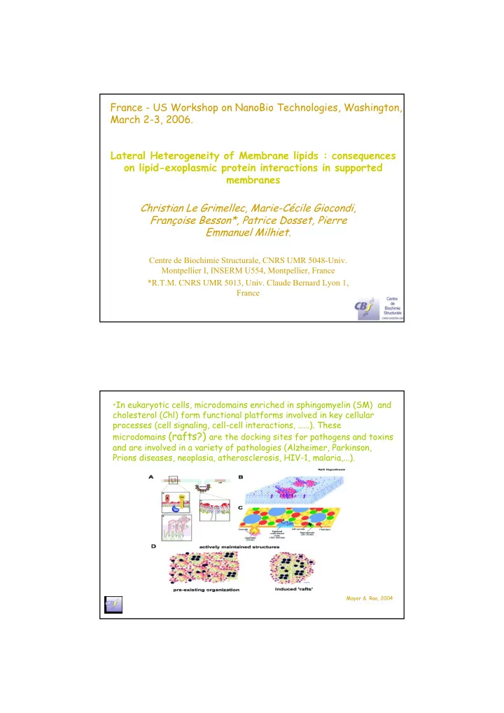
  • In eukaryotic cells, microdomains enriched in sphingomyelin (SM) and

cholesterol (Chl) form functional platforms involved in key cellular processes (cell signaling, cell-cell interactions, ……). These microdomains (rafts?) are the docking sites for pathogens and toxins and are involved in a variety of pathologies (Alzheimer, Parkinson, Prions diseases, neoplasia, atherosclerosis, HIV-1, malaria,...).

Mayor & Rao, 2004

slide-2
SLIDE 2

Part of these functions ( hydrolases, cell adhesion molecules, transmembrane signaling) is performed by exoplasmic proteins linked to the membrane via a glycosylphosphatidylinositol (GPI) anchor. Formation of microdomains would arise primarily from lipid-lipid interactions: lipids constituting rafts are ordered (Lo, Lβ’?) and phase-separate from the disordered (Lα), more fluid species

Goal:

  • To understand the role of lipid-lipid and

lipid-protein interactions in the self- assembling processes of functional platforms.

slide-3
SLIDE 3

g f lo g

Bilayers under buffer

Presumed size of domains suggests a mesoscopic approach: Atomic force microscopy on supported bilayers.

MICA bilayer fusion MICA SUV

buffer

Atomic Force Microscope

(Binnig, Quate, & Gerber. P.R.L., 1986)

Lipid-lipid interactions

slide-4
SLIDE 4

DPPC POPC

Cholesterol

SM

Tc=20°-45°C Tc=41°C Tc=-(2°)C

DOPC

Tc=-20°C

Polymorphism of sphingomyelin microdomains

DOPC/SM (1:1) Existence of globular/ripple structures and gel/gel ϕ separation bar:1 µm

slide-5
SLIDE 5

Details of globular/ripple structures

bar:500 nm Polymorphism of sphingomyelin microdomains

DOPC/SM (1:1)

Polymorphism of sphingomyelin microdomains

POPC/SM (1:1) Details of globular/ripple structures bar:500 nm

slide-6
SLIDE 6

Topography of cholesterol containing bilayers: DOPC/SM/Chl bilayers

  • 2-D « egg-carton » pattern, Chl- dependent
  • is not induced by hypertonic sucrose
  • reproducible shape and periodicity

(~215 nm) between different samples. Salt-induced SM polymorphism: effect of hypertonic NaCl solution

  • n POPC/SM/Chl
slide-7
SLIDE 7

What does the mesoscopic view of supported bilayer tell us about the behavior of functional platforms lipidic constituents?

  • Polymorphism of individual SM microdomains in plasma

membrane models (different organizations energetically close).

  • Reversible changes in microdomains organization can be

induced by limited modification of the medium. Do lipid composition and polymorphism of model microdomains affect the distribution of a GPI- anchored protein (Alkaline Phosphatase from bovine intestine, APase) within the bilayer?

  • Direct insertion of APase in preformed bilayers.

Lipid-Exoplasmic GPI-anchored protein interactions

Protein - C O - NH -(CH2)2 - P GlcNH2 Inositol (Man)3 P (CH2)

16

CH3 CO CH2 - CH - CH2 (CH2)

14

CH3 CO

slide-8
SLIDE 8

APase spontaneously inserts in the lipid ordered (Lβ ’) gel phase, preferentially at the periphery. Moderate increase in the ordered domains area. (Room temperature). Long range order in the APase distribution.

DOPC/DPPC bilayers

bar: 2 µm A,B,C 0.5 µm D z= 10 nm A & B, 5 nm D

APase spontaneously inserts in the lipid ordered (Lβ’) gel phase . APase accumulates at the gel-fluid interface but distributes homogeneously in the gel phase. Remodeling but practically no change in ordered domains area. (Room temperature)

DOPC/SM

bar: 2 µm z=15 nm

slide-9
SLIDE 9

APase preferentially localizes in the thickest SM subdomains (highest Tc?) whose area is markedly increased. Heterogeneity in the APase distribution in the other gel regions ( gel phase physical state heterognenity?) Polymorphism of DOPC/SM samples

bar: 4 µm A-C, 1.25 µm D. Z=15 nm A-C, 10 nm D.

40 µm 30 µm 15 µm

APase localizes in particular regions of the fluid/gel interface. These regions are most often induced by the interaction of the protein with SM domains.

POPC/SM (1:1)

+ APase

slide-10
SLIDE 10

APase recruits the most ordered SM species in its environment.

POPC/SM (1:1)

bar: 4 µm A-C; 1.6µm D; 0.5 µm E z=15 nm A-D; 12 nm E

Distribution of APase in the ordered domains varies according to the lipid species constituting both the ordered (gel) and fluid

  • phases. Conversely, APase induces a remodeling of gel domains most

likely by recruiting the highest melting temperature lipid species.

Effect of cholesterol on APase distribution?

slide-11
SLIDE 11

Addition of 15 mol% Chl often results in the branching of lo-enriched

  • domains. APase distributes homogeneously in the ordered phase. (room

temperature)

DOPC/SM + 15 mol% Chl + APase

(room temperature)

POPC/SM/Chl (1:1:0.35)

+ APase

25°C

A:In contrast to DOPC /SM/Chl bilayersAPase localizes into domains which do not exactly superimposed with the

  • rdered (Lo?) domains.

B: Using 300 pN scan force sweeps away most of the APase. The domains left are thicker and more irregular than the Lo ’s. More ordered (Gel phase) domains?

A B

slide-12
SLIDE 12

18°C 28°C 34°C

POPC/SM/Chl (1:1:0.35)

For low APase/lipid ratio temperature- dependent experiments support the hypothesis of a gel phase environment for the APase.

Almeida et al., 2004

POPC/SM/Chl (1:1:0.35) + high amount APase

At low temperature: APase localizes in SM enriched domains This localization in SM domains is maintained through the macro-ripple stage. At a higher temperature, APase localizes in both SM and POPC enriched domains.

slide-13
SLIDE 13
  • The degree of PC unsaturation affects the distribution of

APase between PC/SM domains both in the absence and in the presence of cholesterol.

  • APase distribution is markedly affected by the temperature

(i.e. the physical state) of the sample.

  • APase preference for the most ordered regions.
  • APAse contributes to the selection of its environment which

is made of the most ordered SM species.

Membrane distribution of the APase

Conclusions

  • Unidirectional direct insertion of an exoplasmic GPI-

anchored protein into a pre-formed supported bilayer under phase-separation.

  • Self-assembling properties upon insertion of a second

exoplasmic protein ?

slide-14
SLIDE 14

V.Vié, C. Domec, O. Bagdhadi, T. Plénat, S. Boichot, CBS, Montpellier

  • D. Lévy, J-L. Rigaud, Institut Curie, Paris
  • E. Lesniewska, J-P.Goudonnet, Laboratoire de Physique,

Université de Bourgogne, Dijon. B.Roux ,F. Ronzon, F. Besson, Université Claude Bernard, Lyon

  • N. van Mau, F. Heitz, S. Deshayes

CRBM, Montpellier

  • M. Zinke-Allmang, Dept. Physics & Astronomy,

University of WesternOntario, Canada.

slide-15
SLIDE 15
  • The degree of PC unsaturation affects the distribution of

APase between PC/SM domains both in the absence and in the presence of cholesterol.

  • APase distribution is markedly affected by the temperature

(i.e. the physical state) of the sample.

  • APase preference for the most ordered regions.
  • APAse contributes to the selection of its environment which

is made of the most ordered SM species.

Membrane distribution of the APase

Conclusions

  • Unidirectional direct insertion of an exoplasmic GPI-

anchored protein into a pre-formed supported bilayer under phase-separation.

  • Self-assembling properties upon insertion of a second

exoplasmic protein ?