first of all what is the definition of the term
play

First of all, what is the definition of the term microfossils? Dr. - PDF document

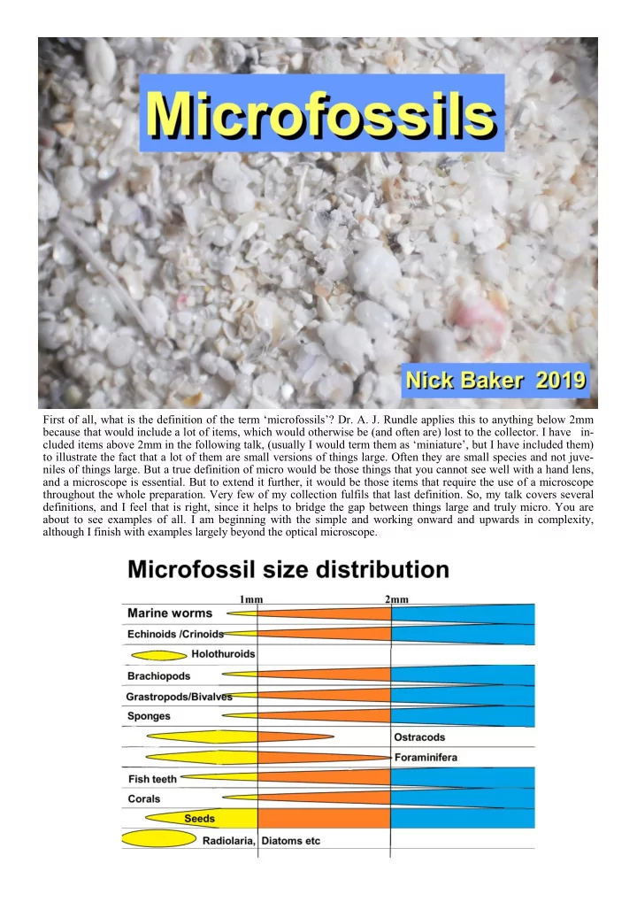
First of all, what is the definition of the term microfossils? Dr. A. J. Rundle applies this to anything below 2mm because that would include a lot of items, which would otherwise be (and often are) lost to the collector. I have in- cluded


  1. First of all, what is the definition of the term ‘microfossils’? Dr. A. J. Rundle applies this to anything below 2mm because that would include a lot of items, which would otherwise be (and often are) lost to the collector. I have in- cluded items above 2mm in the following talk, (usually I would term them as ‘miniature’, but I have included them) to illustrate the fact that a lot of them are small versions of things large. Often they are small species and not juve- niles of things large. But a true definition of micro would be those things that you cannot see well with a hand lens, and a microscope is essential. But to extend it further, it would be those items that require the use of a microscope throughout the whole preparation. Very few of my collection fulfils that last definition. So, my talk covers several definitions, and I feel that is right, since it helps to bridge the gap between things large and truly micro. You are about to see examples of all. I am beginning with the simple and working onward and upwards in complexity, although I finish with examples largely beyond the optical microscope.

  2. Foraminifera . Foraminifera are protozoa. They are largely marine. Mostly single cells. They do not form tissues but consist entirely of protoplasm. My first photo demonstrates the fact that their remains can constitute whole beaches of foraminifera sands. My first single example is of Nummulites . This from Eocene limestone at Al Jabal al Akhbar, Libya. This genus includes the largest of the Forams by a large margin if we include the Nummulites Limestones at Giza. The picture is of a thin section and the cut has been across the examples in those in the lower part of the picture, where the cut is along the line of the cells, while those in the upper part are cut across the speci- men. The sediment appears to have been disturbed. The examples of Globigerina (below) are of a planktonic type, but are not the easiest to find, due to their smaller size. These are from the Upper Chalk at Boxley. Those that are most apparent in any search are the Lenticulinids, (below) and range up to 3mm. These are from the Upper Chalk at Blue Bell Hill. Variations on that form are the “ Ammonia ”, (right) which look like micro - ammonites, and a compart- ment formation can be seen. These are from the Gault Clay at Eccles, Kent. In the case of Ammobaculites (below) the outer shell is thick, but again shows the chambers. The shell is of compact calcite, otherwise it would not survive the preparation processes. The juvenile clump of chambers is fol- lowed by straight construction. In Rectobolivina the chambers surround a central duct, while in Frondicularia the chambers form a plate - like construction. See next page

  3. Sponges Sponges are normally found as large fossils, and in smaller form are included in what I would class as ‘miniature’, as in the example shown to the right, (so far not identified) from the Upper Chalk at Cuxton. Porosphaera are common in the Upper Chalk and range up to 2cm in diameter. Large, hand - size flint nodules are not Porosphaera but are hand - size flint nodules. Porosphaera are calcareous and do not metabolize silica. In thin section (below), the tubular structure is clearly shown in the calcite construction. Flint cores can be mis- taken for Porosphaera but usually show a maze - like chal- cedony in section. Also, flint cores have a rough surface, while Porosphaera shows evenly - spaced micro - pores and have a duct leading to the interior. Porosphaera can be converted to silica on burial but will still show some of the original structure. Porosphaera can range down to 1mm diameter, Sponge spicules are generally found inside nodular flints. I have never found them in Chalk samples. The slightly raised pH in the Chalk environment may render small silica structures soluble. The lower pH in the decay environment in the flint (burial chamber) might prevent this. The spicules are often tubules, forming the circulatory system, as well as support in the sponge structure. The type of spicule is often indicative of the genus to which the deceased belonged.

  4. Bryozoa Bryozoa are colonial animals, (almost all marine) each of the apertures indicating the position of an individual animal. They appear first in the Ordovi- cian, although all the Palaeozoic forms are now extinct. The Mesozoic survivors reached their ‘high noon’ in the Upper Cretaceous. The term Bryozoa (branched - life) covers several orders and sub - orders. In some thin beds of the Upper Chalk they comprise a large percentage of the chalk, almost comprising a ‘reef’. Most are branch - like but they are often found as flat plates, sometimes covering other organisms, earning them the name of ‘sea - mats’ This encrustation can been seen here. Herpetopora anglica can be seen encrusting a bivalve fragment, in the Upper Chalk at Upper Halling, Kent I mentioned the term ‘reef’. This is certainly the case in the Upper Permian, Magnesian Limestone. This is a thin section through a sample from the reef base in the Tunstall Hills, County Durham. One is reminded that the term Coralline Crag of the Pliocene of Suffolk, refers to Bryozoa and not Corals. Corals Corals do not often occur as microfossils – save per- haps fragments in coralline sands. But here I want to refer to the smallest I have found. As a hand - sample, Micrabacia coronula is not uncommon in the Lower Chalk and range up in size to 10mm. Here (above) is an example from Blue Bell Hill. In South Devon, the Lower Chalk is represented by a few metres of hard Cenomanian Limestone. The Hooken Limestone Division below Seaton Cliffs gave the (2mm) example of Micrabacia coronula (left).

  5. Next is a thin section (right) through Devonian lime- stone, from the stone quarry at Ferques, in the Pas de Calais. Here the coral colonies are well shown, the cells being infilled with calcite. I have not been able to name the species. Archaeocyathus I include Archaeocyathus (Ancient Cups) because it is considered to represent the extinction of possibly a whole class. They appear to have been colonial but they differ from Corals, or Bryozoa. They appear in the Lower Cam- brian and are extinct by the Mid Cambrian. This is a thin section through an example from Copley, South Australia. Worms Worms are relatively rare in processed samples, due to the fact that they do not always produce shells. At some levels in the Chalk they are quite abundant as marine worms. Some are organised and ornamented. Some are unorna- mented and apparently disorganised in their growth, with Glomerulus as a good example of the latter in the Upper Chalk. They have been likened in appearance to squirts of polyfiller. There are two examples shown here Others, such as Neomimicrorbis have strong ornamentation. What the environmental advantages are in these two situations is difficult to say. This specimen is from the Upper Chalk at Meresborough, near Rainham, in Kent

  6. Echinoids We are now going to look at Echinoderms, which com- prise Echinoids, Crinoids, Asteroids, and Holothuroids. So for Echinoids, in the Chalk, the most common remains are the plates or fragments of the test. By some referred to as the rivets (right) And here is a close - up (below) of one such, probably from a Cidarid . Other common fragments are the spines. And also fragments of the ambulacra, part of the respire - tory system. (below). Very occasionally it is possible to find juvenile examples of Echinoids, mainly Cidarids. And here are two exam- ples of such. It has been pointed out the five - fold struc- ture is not possible to see in these. The answer is that such development may become apparent only in the adults. The infant state of an Echinoid is also very short compared with the rest of its life - span Other items to be found are the ‘lanterns’ (right). These form part of the ‘teeth’ in the mouth, largely used for rasp- ing, rather than biting. The area is quite complex but goes back to the earliest stages of Echinoid evolution in the Ordovician.

  7. Cidarids At the small level the most common remains of Crinoids are stem fragments. Here is a selection. And an example (below) from the Lower Chalk at Blue Bell Hill. It is possible that fragments of calyx may be common but may be less easily noticed. Holothuroids Holothuroids (see below) include sea - cucumbers. The wheel - like structures here, consist of part of the supporting ‘skeleton’ which is here about 250 microns across, and were found in the Jurassic Oxford Clay, near Weymouth, Dorset. Brachiopods Brachiopods were the most common in the Palaeozoic, but retained their abun- dance in the Mesozoic. They are much depleted in the Tertiary, giving way to more modern bivalves. In the Middle Chalk small Rhychonelids are abun- dant, especially Terebratulina of which we see several here. One can see why they are sometimes referred to as ‘lamp - shells’. But of special note, I want to illustrate Iso - Crania , (below) from the Upper Chalk, where the posterior adductor muscles are located well to- wards the centre of the valves. Likewise with the exterior ridges. The effect is to award the name of ‘chicken face’. One is not usually rewarded with a view of the interior. The ornamented surface tends to accommodate ‘resis - tant’ chalk and cleaning small fossils is difficult and risky.

Download Presentation
Download Policy: The content available on the website is offered to you 'AS IS' for your personal information and use only. It cannot be commercialized, licensed, or distributed on other websites without prior consent from the author. To download a presentation, simply click this link. If you encounter any difficulties during the download process, it's possible that the publisher has removed the file from their server.

Recommend


More recommend