
SLIDE 1
Feasibility Study of X-ray Fluorescence Imaging System: Surface Modification Gold Nanoparticles and 2D Convolutional Neural Network
Taeyun Kim a,c, Wooseung Lee a, Jimin Lee a, Sung-Joon Ye a,b*
aProgram in Biomedical Radiation Sciences, Department of Transdisciplinary Studies, Graduate School of
Convergence Science and Technology, Seoul National University, 08826, Seoul, Republic of Korea
bAdvanced Institutes of Convergence Technology, Seoul National University, 16229, Suwon, Republic of Korea cNeutron and Radioisotope Application Research Division, Korea Atomic Energy Research Institute, 34057, Daejeon,
Republic of Korea
*Corresponding author: sye@snu.ac.kr
- 1. Introduction
Recently, researches on tumor diagnosis and radiation therapy using metal nanoparticles (MNPs) (e.g., gadolinium, platinum, silver, and gold) have been actively conducted. Among them, gold nanoparticles (GNPs) have been most extensively studied. Because gold is an inert metal, its biocompatibility is superior to
- ther MNPs. Moreover, various types of surface
modifications (e.g., poly(ethylene) glycol, antibody, and protein) can be used to improve tumor-targeting ability (e.g., enhanced permeability and retention (EPR) effect). After GNPs accumulated in the tumor region, they can serve as the imaging contrast agents and radiosensitizers due to the high X-ray absorption coefficient derived from a high-atomic number [1]. When conducting in vivo imaging and therapeutic studies with MNPs, it is very important to evaluate the biological effects. For a more accurate evaluation, it is essential to obtain the in vivo biodistribution of MNPs. X-ray fluorescence computed tomography (XFCT) and X-ray fluorescence (XRF) imaging based on X-ray fluorescence (i.e., Characteristic X-ray) are emerging as promising in vivo imaging modalities that can obtain the in vivo biodistribution of MNPs (i.e., XRF image) [2, 3]. In 2019, a benchtop XFCT system with a linear array cadmium zinc telluride (CZT) detector and a single pinhole collimator was developed for in vivo imaging of the gadolinium nanoparticles in living mice [2]. This benchtop XFCT system required an XRF image acquisition time of 7.5 min per target slice. More recently,
- ur previous study reported an XRF imaging system with
a commercial 2D CZT gamma camera and a single pinhole collimator for dynamic in vivo imaging of the GNPs in living mice [3]. With the implementation of a 2D CZT gamma camera, the XRF image acquisition time was reduced to 2 min per target slice. The aforementioned XFCT and XRF imaging systems have limitations in common with the advantages and disadvantages that are offset by each other. In the case of the XRF imaging system, the dynamic in vivo XRF images could be obtained due to dramatically shorter image acquisition time than the XFCT system. Whereas, the XRF imaging system has a limitation that essentially requires an XRF image before the injection of MNPs (i.e., pre-scanning) for the direct subtraction method (i.e., Compton background elimination), unlike the XFCT
- system. This can generate artifacts by position error and
double the imaging dose and image acquisition time. In addition, both systems investigated the feasibility of in vivo XRF image acquisition using pure MNPs without surface modification. In this study, we investigated the feasibility of XRF imaging system with surface modification GNPs and 2D convolutional neural network (CNN) for Compton background elimination. The in vivo XRF image
- btained by injecting liposomal GNPs into tumor-
bearing mice was used to investigate the tumor-targeting ability improvement of liposomal GNPs. Furthermore, for substituting pre-scanning XRF image acquisition, which was the major limitation of our previous XRF imaging system, Compton background elimination was performed using pre-developed 2D CNN.
- 2. Methods and Results
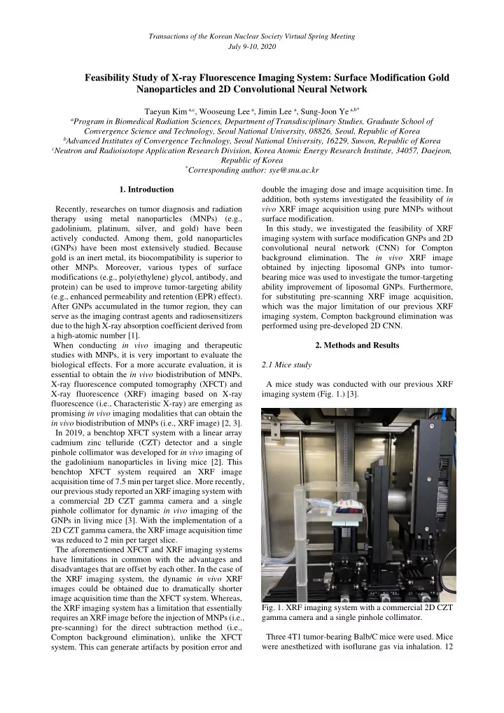
2.1 Mice study A mice study was conducted with our previous XRF imaging system (Fig. 1.) [3].
- Fig. 1. XRF imaging system with a commercial 2D CZT