SLIDE 12 +
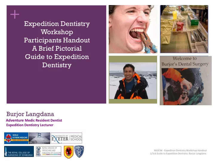
NSOCM - Expedition Dentistry Workshop Handout 1/3-A Guide to Expedition Dentistry- Burjor Langdana.
Is there need for Pre-departure Dental Check Up? Answer- It'll help in the Prevention and Diagnosis of Teeth andGum problems on an Expedition.
4.
A) Pre Departure Dental Check ups- Prevention- Pro Active involvement in making sure this is done at least
2/3 months before departure. Informing the dentist where, how long for and degree of dental cover available. i) 2/3 months gives enough time for the dentist to complete complicated treatment like Root Canal, Crowns, Extractions. 2/3 months also affords sufficient time to heal from these procedures. ii)Informing the Dentist about duration, location and degree of Expedition Dental cover. Allows him to adjust his treatment accordingly. Especially for those teeth that lie in the grey zone between conservative and radical treatment modalities 1 2 3 4 5 6
B)Pre Departure Dental Check ups- Diagnosis- To collect a Detailed Dental Charting from the Dentist and
Carry it on the Expedition. To an untrained eye the tooth may look perfect. But actually it could be a crown, a white composite filling. All these may just look like a tooth. A detailed dental charting will let you know exactly Where are the= Fillings: Amalgam and White composites Where are the = Crowns Where are the problem areas= Deep Fillings Where are the=Root Canal treated Teeth Where are the= Wisdom Teeth, are they Impacted, or have they been removed 3 4 5 6