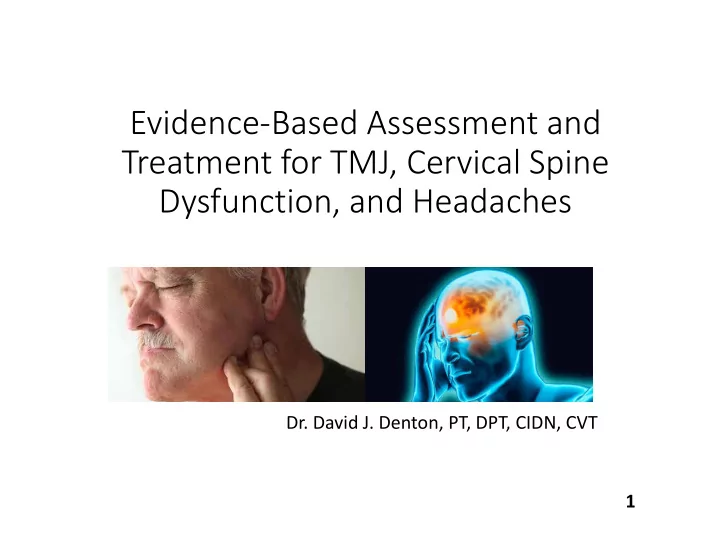
SLIDE 1 Evidence‐Based Assessment and Treatment for TMJ, Cervical Spine Dysfunction, and Headaches
- Dr. David J. Denton, PT, DPT, CIDN, CVT
1
SLIDE 2 Learning Objectives
- Utilize your client’s functionality through relief of neck and jaw
pain through manual based intervention for immediate relief.
- Examine clinical practice and review differential diagnosis, cervical
spine, balance system, and jaw physiology/anatomy, and symptomatic connection.
- Establish the best practices in assessment and re‐assessment
strategies for relieving pain, underlying conditions, and manual based lab techniques.
- Demonstrate different treatments, exercise prescription, and
documentation for TMD patients.
- Participate in interactive hands‐on labs for assessing the
involvement of the cervical spine (cervicogenic vertigo, cervicogenic headache), affected balance, and TMD treatment approaches.
- Implement effective , evidence based interventions for all
involvement at the upper cervical spine resolving dizziness, restoring the balance system, resolving headaches, and relief of TMD pain. 2
SLIDE 3 Cour Course se Outline Outline
- 8:00 Connection of Cervical Spine, Headaches, and TMD
- 8:30 Upper Cervical Spine, Headaches, and Upper Cervical Nerves
- 9:00 First Rib, Thoracic Outlet, and Radicular Symptoms
- 9:15 Upper Cervical Palpation Lab
- 10:00 BREAK
- 10:15 Describe causation of balance disorders and define vestibular rehab.
- 10:30 Enhance knowledge base of sensory and motor control roles.
- 10:45 Postural Control Lab
- 11:30 Upper Quarter Screen/Special Tests and Clearing Exam with Lab
- 12:00 Lunch
- 1:00Cervicogenic Dizziness and Vestibular Hypofunction
- 1:30 CGH Treatment theories and Exercise prescription.
- 2:00Vestibular Rehabilitation, Cervicogenic Vertigo and Hypofunction Lab
- 2:45 Break
- 3:00 Biomechanics and Pathology of TMD
- 3:30 TMD Evaluation (Videos and Lab) and Treatments
- 4:15 Comments, Course Evaluation, and Closure
3
SLIDE 4 Connection of Cervical Spine, Headaches, and TMD
- What does the evidence say?
- TMD ‐ commonly associated with conditions of the
head and neck region.
- Cervical spine disorders (including Cervicogenic
Dizziness)and Headache.
- Presence of neck pain was shown to be associated with
TMD 70% of the time. (Silveira, 2015)
4
SLIDE 5 Cervical Connections
- Most of the studies agree that symptoms from the
cervical spine can be referred to the stomatognathic (teeth, jaws, tissue) region through the trigeminocervical nucleus.
- Presence of signs and symptoms in the cervical region
- f patients suffering with TMD and that the presence of
tender points in the cervical area of these patients co‐ exist.
- Muscle tenderness in the cervical spine and jaw was
shown to be associated with increased levels of jaw and neck disability (Silveira, 2015).
5
SLIDE 6 Whole Body Connection?
- The body as a whole operates on the principle of
- compensation. Disturbances in the upper quarter like
increased muscle tension leads to compensatory changes within the muscle tension in the spinal region.
- Review of the literature showed that pain in the upper
quarter and masticatory motor system may be caused by cervical spine disorders (generally by dysfunction of muscular origin) and vice versa (Dragon, 2014).
6
SLIDE 7 Cervical Spine and Headaches
- Types of Headaches:
- Primary headaches include those of vascular origin
(cluster and migraine headaches) as well as those of muscular origin (tension‐type headaches).
- Secondary headaches result from another source
including inflammation or head and neck injuries (Cervicogenic)
7
SLIDE 8 Upper Cervical Spine and Headache Connection
- Criteria for Neck Pain and Headache:
- Unilateral pain with a facet ‘lock’ irradiating from the
back of the head
- Evidence of cervical dysfunction presenting during
manual examination
- May occur with trigger point palpation in the head or
neck
- Aggravated by sustained neck positions
- Normal imaging (Page, 2011)
8
SLIDE 9 Upper Cervical Spine and Headache Connection
- Trigeminal Pathway:
- It is suggested that CGH is a “final common pathway” for
pain generating disorders of the neck.
- Proposed pathophysiology theory: CGH results from a
convergence of sensory input from the upper cervical spine into the trigeminal spinal nucleus, including input from:
- Upper cervical facets
- Upper cervical muscles
- C2‐3 intervertebral disc
- Vertebral and internal carotid arteries
- Dura mater of the upper spinal cord
- Posterior cranial fossa (Bogduk, 2014).
9
SLIDE 10 Headache Statistics
- Approximately 47% of the global population suffers
from a headache.
- 15‐20 percent of those headaches are
- Epidemiological researchers suggest a higher
prevalence of headache in adults with neck pain.
- Females seem more predisposed to CGHs affecting 4
times as many women as men. (Page, 2011).
10
SLIDE 11 Red‐flags Associated with Headache
- Headaches that are getting worse over time
- Sudden onset of severe headache
- Headaches associated with high fever, stiff neck, or rash
- Onset of headache after head injury
- Problems with vision or profound dizziness
11
SLIDE 12 Pathophysiology of Cervical Spine and Headache Screening
- Nearly 50% of CGH patients as well as asymptomatic
subjects had bulging cervical discs.
- The most important clinical finding to diagnose CGH is
impairment of C1‐C2 (atlanto‐axial) motion.
- CGHs can occur in patients with lower cervical dysfunction,
particularly after trauma.
- Preadolescent (10‐13 year old) children with neck pain and
headache have dysfunction of the lower cervical spine.
- Cervical osteoarthritis, common in many older patients may
be associated with headache and cervical muscle
- Diagnosis of CGH should be made by carefully examining the
etiology of the headaches, not just the symptoms.
12
SLIDE 13
Trigeminocervical Nucleus
13
TCN Trigeminal Nerve TCN – Trigeminocervical Nucleus C1‐3 – Spinal Nerve Roots of C1 to C3 C1 C2 C3
SLIDE 14 Diagnostic Criteria
14
Major criteria
- I. Symptoms and signs of neck involvement
- a) Precipitation of comparable symptoms by:
- 1) neck movement and/or sustained,
awkward head positioning, and/or
- 2) external pressure over the upper cervical
- r occipital region
- b) Restriction of range of motion in the neck
- c) Ipsilateral neck, shoulder or arm pain
- II. Confirmatory evidence by diagnostic anaesthetic block
- III. Unilaterality of the head pain, without sideshift
Head pain characteristics
- IV. Moderate‐severe, non‐throbbing pain, usually starting
in the neck
- Episodes of varying duration, or fuctuating, continuous
pain
SLIDE 15 Migraine vs. CGH
15
Migraine CGH
Gender ratio 1.69 female/male 0.71 female/male Age at onset 18 years 33 years Headache onset Anterior head Posterior head/neck Pain area 50% unilateral Predominately unilateral Nausea Frequent Infrequent Photo/phonophobia Very frequent Infrequent T robbing pain Frequent Infrequent Pain increases when bending forward Very frequent Infrequent Migraine medication Usually helpful Not helpful Sustained/awkward neck position provokes pain Rare Universal
SLIDE 16 TMJ and CGH Connection
- Temporomandibular Disorders (TMDs) and
headache are closely related pathologies; prevalence of headache in the dysfunctional population varies between 48% and 77%, while in the general population the prevalence of headache is around 45% (Paolo, 2017).
16
SLIDE 17 TMJ and CGH Connection
- According to several studies, there is a strong
correlation between headache and other dysfunctional symptoms, such as joint noise, pain during mandibular movement, pain in the temporomandibular area, depression, anxiety, and poor sleep quality (Paolo, 2017).
17
SLIDE 18
Trigeminocervical Nucleus
18
SLIDE 19 Occipital Nerves and HA/Dizziness
- Occipital Neuralgia (C2 neuralgia), is a medical condition
characterized by chronic pain in the upper neck, back of the head and behind the eyes. These areas of the head correspond to two nerves located in the back of the neck at the base of the skull.
- Greater and Lesser Occipital nerves.
- Symptoms of the condition:
- chronic headache, tenderness and pain at the base of the skull, in
- ne or both sides, aching, burning, and throbbing pain that
typically starts at the base of the head and radiates upwards into the scalp, pain behind the eye, sensitivity to light and tenderness throughout the scalp.
- Causes of Occipital Neuralgia:
- compression to either the greater or lesser occipital nerves as they
penetrate the muscles high up in the neck just under the skull lateral to C2.
- Secondary to whiplash, or poor posture and head position that
develops gradually over time. 19
SLIDE 20 Auriculotemporal Nerve
- The superficial temporal branches run with the
superficial temporal arteries.
- Branches innervate the skin over the temple and
anastomose with the facial and also the zygomaticotemporal nerves.
- Branches to the external auditory meatus, which are
typically two in number, run between the cartilaginous and bony ear canals and innervate the skin of the meatus, but also provide innervation to the tympanic membrane.
20
SLIDE 21 Greater Auricular Nerve
- The great auricular nerve (or greater auricular nerve)
- riginates from the cervical plexus, composed of branches of
spinal nerves C2 and C3.
- It provides sensory innervation for the skin over parotid
gland and mastoid process, and both surfaces of the outer ear.
- It can be damaged by the neck surgery, tumor,and
prolonged pressure on the neck.
- Can be an etiology of parietal pain and headaches.
21
SLIDE 22 Current Perspectives on Orofacial Pain
- Innervation of trigeminal system (head, face, masticatory
musculature, temporomandibular joint and associated structures).
- Temporomandibular disorders (TMD) are the most prevalent
- rofacial pain conditions for which patients seek treatment.
- Temporomandibular disorders include a number of clinical
problems that involve the masticatory musculature, the temporomandibular joint (TMJ) or both.
- Trigeminal neuropathic pain conditions can arise from injury
secondary to dental procedures, infection, neoplasias, or disease or dysfunction of the cervical spine (Reyes, 2015).
22
SLIDE 23 The First Rib and Pain Referral
- First Rib ‐ most superior of the twelve ribs.
- Atypical rib and anatomical landmark.
- It is one of the borders of the superior thoracic aperture
- Innervation
- Innervated by the first intercostal nerve.
- The intercostal nerves are distributed chiefly to the
thoracic pleura and abdominal peritoneum and differ from the anterior rami of the other spinal nerves in that each pursues an independent course without plexus formation.
23
SLIDE 24
24
SLIDE 25 The First Rib and Pain Referral
- So Many Attachments!!
- Anterior scalene muscle: scalene tubercle
- Middle scalene muscle: between groove for the
subclavian artery and transverse tubercle
- Intercostal muscles: from the outer border
- Subclavius muscle: arises from the distal shaft and first
costal cartilage
- First digitation of the serratus anterior muscle
- Parietal pleura: from the inner border
- Costoclavicular ligament: anterior to the groove for
the subclavian vein
25
SLIDE 26 How to Palpate the First Rib
- The first rib is often noted as the most difficult rib to
- palpate. To palpate the first rib, find the superior border
- f the upper trapezius muscle and then drop off it
anteriorly and direct your palpatory pressure inferiorly against the first rib. Asking a patient to take in a deep breath will elevate the first rib up against your palpating fingers and make palpation easier
- https://youtu.be/zwDO_VXOItM
- https://youtu.be/PrDZD1erucI
26
SLIDE 27 First Rib Pathology
- Thoracic Outlet Syndrome ‐ compression of the
neurovascular structures as they exit through the thoracic outlet (cervicothoracobrachial region)
- The thoracic outlet is marked by the anterior
scalene muscle anteriorly, the middle scalene posteriorly, and the first rib inferiorly.
- The term ‘TOS’ does not specify the structure being
compressed.
27
SLIDE 28 First Rib Pathology
- TOS affects approximately 8% of the population and is
3‐4 times as frequent In woman as in men between the age of 20 and 50 years.
- Signs and symptoms of thoracic outlet syndrome vary
from patient to patient due to the location of nerve and/or vessel involvement. Symptoms range from mild pain (referred to the shoulder blade/chest and around shoulder) and sensory changes to limb threatening complications in severe cases
28
SLIDE 29 First Rib Pathology
- Pancoast Tumor ‐ refers to a relatively uncommon
situation where a primary bronchogenic carcinoma arises in the lung apex at the superior pulmonary sulcus and invades the surrounding soft tissues.
- The most common symptoms at presentation are
chest and/or shoulder pain, with arm pain being also common. Weight loss is frequently present
29
SLIDE 30 TOS Exercises
- https://youtu.be/lnaxIFP2_yA
- https://youtu.be/q0r5XfZ‐NXc
- https://youtu.be/MpojqL‐B5pc
- https://youtu.be/wz6KRl3YtqU
- https://youtu.be/YQMpmJbpfgY
30
SLIDE 31 TOS Exercises
- Literature synopsis of first rib mobilization:
- While literature investigating manual therapy in the
treatment of thoracic outlet syndrome is very limited, results show a manual depression of the 1st rib to be an easy, effective option in conservative management
- f thoracic outlet symptoms.
31
SLIDE 32 Causa Causation ion of
Balance Di Disor sorder ers and and Vestibular ibular Rehabilit habilitation ion De Defined ned
- Function
- Statistics
- Etiology
- Gait Assessment
- Balance Testing
32
SLIDE 33 Function of the Vestibular System
- The vestibular system functions:
- Detects motion of the head and maintains stability of images on
the fovea of the retina
- Vestibular‐ocular reflex
- postural control during head motion (cervical), ‐Minor 1998
- Vestibulo‐spinal reflex
- The system is made up of 3 components:
- Peripheral sensory apparatus (Picture next Slide)
- Motion sensors that send info to the CNS (Vestibular nucleus complex
and cerebellum)
- Information on head velocity and linear acceleration
- Central processor
- The CNS
- Processes signals to estimate head and body orientation in space
- A mechanism for motor output
- Info is sent to ocular muscles and the spinal cord in 3 reflexes:
- Vestibulo‐ocular reflex (VOR)
- Vestibulocollic reflex (VCR)
- Vestibulospinal reflex (VSR)
33
SLIDE 35 Function of the Vestibular System
- VOR – generates eye movements
- Enables clear vision while the head is in motion.
- Stabilizes images on the retina
- Elicits eye movement by stimulating the vestibular system
(Nystagmus)
- Nystagmus is rhythmic back and forth beating (VOR generates
slow phase to keep eye focused)
- When eye reaches max range; a Saccade (quick phase) in
generated in Opposite direction to new target
- VCR – acts on the neck musculature to stabilize the head
- Reflex initiated by vestibular system. “Righting reflex”
- VSR – generates compensatory body movement to maintain head and
postural stability
- Fall prevention and balance strategies
- The performance of all 3 reflexes is monitored by the CNS and re‐
adjusted accordingly by the cerebellum. – (Hain, T)
35
SLIDE 36 Function of the Vestibular System
- Statistics
- Population based study in US reported that 24‐30% of older
people >age 72 have dizziness (Iwasaki/Klaus)
- Falls are the leading cause of hospital admission and accidental
death in elderly population (Iwasaki)
- Vertigo and unsteadiness causes a fear of falling.
- This is a predictor for those who will suffer one or more
subsequent falls.
- Vestibular rehab significantly decreases the risk of falls in the
elderly (Macias)
- Age related decline of the vestibular system correlates with age
related decrease in the number of vestibular hair cells and neurons (Iwasaki)
- Mitochondrial anti‐ox (alpha lipoic acid and co Q10) may
reduce age dependent hair cell loss in study (Iwasaki 2015)
36
SLIDE 37 Sensory and Motor Control Roles
- Postural Control
- Vestibulopathy,
- Nystagmus Assessment
- Involuntary movement of the
eye
- Balance Components
- VOR – reflex where activation
- f Vestibular system causes
eye movement
- Bilateral vs. Central Loss
37
SLIDE 38 Po Postural Con Control
- Postural control is the ability to control the body position
in space. –Huang 2006
- Normal CNS involvement keeps the body in vertical
postural alignment
- Patients with affected vestibular function will lean, tilt, or
shift toward the weaker side.
- Patients will correct this with compensation strategies.
- Patients immediately experience hypermetria (movement of
a body part beyond its goal) after a vestibular hit
- Ataxic with severe postural instability
- Increased amplitude of both reactive and anticipatory
postural responses
38
SLIDE 39 Postur ural al Con Control
- Boundaries = Limits of stability.
- Ankle strategy allows ~4 degrees backward and ~8 degrees
forward on stable surface.
- In vestibulopathy, responsive reactions are larger
movements than are needed
- Theory for vestibular hypermetria:
- Reduced cerebellar inhibition of the spinal motor system
- Loss of vestibular input reduces the inhibitory mechanism of
Purkinje cells (in cerebellum)
- Poor reactive synapses from the somatosensory system to the
vestibular nucleus after loss of vestibular drive –Hurak 2009 39
SLIDE 40 Po Postural Con Control
- Changes with age related to sensory systems include:
- Reduced joint position sense
- Reduced visual function
- Decreased vestibular function
- In elderly Shumway‐Cook reported a 40% reduction in lower body
muscle strength compared to young and healthy pt
- Altered muscle response organization
- More frequent co‐activations of antagonist muscles
- Elderly exhibited a decreased ability to react to external
perturbations
- Difficulty in sensory re‐weighting
- Lastly “stance of elderly people is reported to be less stable with
absent or altered proprioceptive, vestibular and visual information.” (Peterka) 40
SLIDE 41 Postur ural al Con Control
information regarding sensory data is congruent.
contribute proportionally to body position in space and movement.
- Sensory situations that are
not congruent are:
surface
- Modification or absence
- f vision
41
SLIDE 42 Po Postural Con Control
- Postural Tests per Clinical Preference/Practice for goals and
return of Postural Control Tracking
- Sensory Organization Test (SOT) – objectively identifies
abnormalities in varying three sensory systems
- Modified Clinical Test of Sensory Interaction (mCTSIB) –
derivative of SOT to provide objective evidence of sensory dysfunction
- Dynamic Visual Acuity (DVA) – changes in visual acuity at head
velocities (visited again in formal Eval Section) Highly reliable for Vestbular hypofunction
- 10‐15 feet away from eye chart
- Stability Evaluation Test (SET) – balance control measures of
postural sway velocity on available test conditions (per clinic)
- Others you may wish to employ after independent study or
clinical opinion:
- Motor Control Test, Adaptation Test, Limits of Stability, Gaze
Stabilization test, Unilateral Stance, Sit to Stand 42
SLIDE 43 Po Postural Contro rol
Control
environments, the CNS relies more on the vestibular system for fall prevention
proprioception
- Results in elderly showed
a decline in amplitude of motor feedback.
LAB – Rehab Tx
43
SLIDE 44 Cer Cervical al Scr Screen een and and Special Special Te Tests
- Vertebral Artery Test
- Cervical sidebend and rotation in slight extension
- Chin on Hand testing to mimic Dix Hallpike positioning for
clearance (picture 1)
- Arms extended test watching for drooping of one arm,
parasthesia, dizziness or nystagmus (picture 2 and 3)
- Red Flags: diaphoresis, dysphasia, dysarthria, drop attacks,
diplopia, high pain
44
SLIDE 45
Special Special Te Tests
45
SLIDE 46 Special Special Te Tests
- Cervical Instability
- Sharp Purser – assessment
- f transverse ligament.
- 1. Forward flex head and
support with hand or elbow, opposite hand on SP of C2.
- 2. Soft end feel, clunk or
reduction of symptoms with shear
- 3.Positive test and ER call.
- The test without head
flexion is also an effective headache SNAG 46
SLIDE 47 Special Special Te Tests
- Ligamentous Stability
- Cranial Axis Lift
- Transverse Ligament
Assessment
- 1. Supine
- 2. Support occiput with
fingers on posterior arch of atlas
- 3. gently lift axis and atlas
while preventing upper cervical flexion
- 4. Negative test = hard end
feel
- 5. Excessive motion or soft
end feel = positive test
- Monitor for parasthesia and
nystagmus 47
SLIDE 48 Special Special Te Tests
- Ligamentous Stability
- Atlas – Axis Shear Test for
Transverse Ligament
hand on C2
- Arms parallel to table
- Press hands toward each
- ther in a shear motion
- Hard/Firm end feel is
negative
48
SLIDE 49 Special Special Te Tests
- Ligamentous Stability
- Alar Ligament
- Find C2 Spinous process
- Passively sidebend the upper
cervical spine
- Passively rotate the upper
cervical spine
- Hard end feel = negative
- Soft end feel or increased
pain/spasm = positive test
49
SLIDE 50 Special Special Te Tests
- Ligamentous Stability
- Tectoral Membrane
- 3 positions:
- Gentle long axis traction in
neutral
flexion with traction
axis
feel/lag = positive test
50
SLIDE 51 Upper Upper Quart Quarter Scr Screen een
- Manual Muscle Testing
- Cervical Spine
- ROM
- Compression
- Distraction
- Sensation
- Dull
- Sharp
- Light Touch
- Deep Tendon Reflex
- Normal
- Brisk
- Absent
- Diminished
- Coordination
- Finger to Nose
- Heel Shin
- Rapid Alternation
- Finger Opposition
51
SLIDE 52 Scr Screening eening Lab Lab
- Vertebral Artery
- Transverse Ligament
- Alar Ligament
- Tectoral Membrane
- UE Screen/ROM/MMT/Neuro
52
SLIDE 53 Ce Cervic icog
ic Dizziness zziness
- Cervicogenic dizziness may be a result of whiplash injury,
- ther forms of cervical spine dysfunction, or spasms in the
cervical muscles. Wrisley, 2000
- The diagnosis of cervicogenic dizziness is characterized by
dizziness and dysequilibrium that is associated with neck pain in patients with cervical pathology
- Cervicogenic dizziness is most often associated with
flexion‐extension injuries and has been reported in patients with severe cervical arthritis, herniated cervical disks, and head trauma. In these patients, complaints of ataxia, unsteadiness of gait, or postural imbalance associated with neck pain, limited neck range of motion,
- r headache predominate, Wrisley 2000
53
SLIDE 54 Cervic icogenic ic Dizziness Dizziness
- Pathophysiology
- With strong connections between the cervical receptors and
balance function, it is believed that injury or pathology of the neck may be associated with a sense of dizziness or disequilibrium, (Wrisley 2000)
- Current theory is that cervicogenic dizziness results from
abnormal input into the vestibular nuclei from the proprioceptors of the upper cervical region. (Wrisley 2000)
- The interconnections between the cervical proprioceptors and
the vestibular nuclei may contribute to a cyclic pattern, where the cervical muscle spasms contribute to dizziness; and dizziness contributes to muscle spasm, although the causal relationship is unclear. (Wrisley 2000)
54
SLIDE 55 Cervic icogenic ic Dizziness Dizziness
- Cervicogenic dizziness is a diagnosis of exclusion
- (ie, the diagnosis is usually based on the elimination of the other
competing diagnoses, such as vestibular or central nervous system pathologies), (Wrisley 2000)
- The neck torsion nystagmus test, or head‐fixed, body‐turned
maneuver is considered by some to identify cervicogenic dizziness.
- Test requires the head of the patient to be stabilized while the
body is rotated underneath.
- Theoretically, the neck proprioceptors are stimulated while the
inner ear structures remain at their resting. Nystagmus is elicited in a positive test.
- Determine if the patient with a chief complaint of dizziness or
vertigo has neck pain, either at rest, with active neck movement, or with palpation of the neck musculature.
- This step is important because, by definition, a diagnosis of
cervicogenic dizziness is excluded in a patient without neck pain. (Wrisley 2000) 55
SLIDE 56 Cervic icogenic ic Dizziness Dizziness
- Manual therapy is recommended treatment for cervicogenic
dizziness directed at decreasing muscle spasms and trigger points of pain in the cervical musculature.
- Treatment for cervicogenic dizziness:
- Manual therapy to decrease the irritation on the cervical
proprioceptors from muscle spasms and trigger points
- Exercises with graded exposure to sensory inputs to improve the
patient's use of vestibular and proprioceptive inputs for balance.
- Eye exercises to improve the function of the vestibular‐ocular
reflex.
- Evidence shows the use of manual therapy/mobilization for
cervicogenic dizziness showed improvement in postural stability, ROM, muscle tenderness and neck pain. Therefore manual therapy combined with vestibular rehab yielded good treatment outcomes (Lystad)
56
SLIDE 57 Vestibular ibular Hypofunction Hypofunction
- Defined as postural instability, visual blurring with head
movement and subjective complaints of dizziness
- Results: Based on Strong evidence of benefit over harm
- Clinicians should offer vestibular rehab to uni and bilateral
vestibular hypofunction with impairments and limitations related to deficit
- Clinicians should NOT include voluntary saccadic or smooth
pursuit eye movements in isolation (without head movement)
- Should be perscribed 3 x per day for gaze stability as at least
- ne component of HEP
- Based on expert opinion ‐ acute or subacute unilateral
hypofunction may need 1 x per week for 3 sessions then HEP
- Chronic unilateral 1x per week 4‐6 visits, Bilateral 1 x for 8‐12 V
- Evidence derived: APTA Neurology section Clinical Practice
Guidelines 57
SLIDE 58
Theories, Theories, In Interv rven entio tions, and and Ex Exer ercise cise Pre Prescription
Vestibular Rehabilitation Therapy
58
SLIDE 59 Section Section Obj Objecti ctives es
- 1. Understand the role and effectiveness of Vestibular
Rehabilitation Therapy (VRT) on Unilateral/Bilateral Peripheral Vestibular Hypofunction.
- 2. Review indications for VRT regarding Peripheral Vestibular
Hypofunction including enhanced gaze stability, enhanced postural stability, improved vertigo, and improved ADL’s.
- 3. Understand and apply interventions of adaptation,
substitution, and habituation.
- 4. Demonstration of progressive treatments of VRT to alleviate
the primary and secondary problems caused by vestibular disorders.
- 5. Demonstration and understanding of causation and manual
treatment for cervical caused dizziness.
- 6. Reference of outcomes and considerations for various
conditions
59
SLIDE 60
60
SLIDE 61
61
SLIDE 62 Ce Cervic icog
ic Dizziness zziness
- Possible result of cervical spine dysfunction including:
- Whiplash injury
- HNP
- Forms of cervical spine dysfunction (Trauma/OA)
- Spasms in the cervical muscles. (Wrisley, 2000)
- Diagnosis
- Characterized by dizziness and dysequilibrium
- Associated with neck pain in patients with cervical pathology
- True spinning vertigo is rarely associated with this syndrome
- Presentation and Symptoms
- Complaints of ataxia/unsteadiness
- Headache
- Postural imbalance associated with neck pain
- Limited neck range of motion (Wrisley 2000)
62
SLIDE 63 Cer Cervic icog
enic Dizziness Dizziness
- Pathophysiology
- Strong connections between the cervical receptors and
balance function
- Injury or pathology of the neck ‐ associated with a sense of
dizziness or disequilibrium, (Wrisley 2000)
- Current theory
- Abnormal input into the vestibular nuclei from the
proprioceptors of the upper cervical region. (Wrisley 2000)
- Cyclic pattern
- Cervical muscle spasms contribute to dizziness
- Dizziness contributes to muscle spasm, although the causal
relationship is unclear. (Wrisley 2000)
63
SLIDE 64 Vestibul ibular ar Hypof Hypofunction unction
- Bilateral Vestibular Hypofunction
- Reduction or loss of vestibular function
- Difficulty walking in the dark or on uneven surfaces
(Balance)
- Decrease in the patient’s ability to see clearly during
head movements.
- Ototoxicity, meningitis, sequential vestibular neuritis,
progressive disorders, autoimmune disorders, and
- neurofibromatosis. (usually idiopathic)
- Oscillopsia ‐ Another problem for patients with
bilateral vestibular loss is the visual blurring that
- ccurs during head movements. Almost 70% of all
patients with BVH complain of oscillopsia even after a course of gaze stabilization exercises.
64
SLIDE 65 Vestibul ibular ar Hypof Hypofunction unction
- Unilateral Vestibular Hypofunction (UVH)
- A condition in which one of the peripheral vestibular
receptors or the vestibular portion of cranial nerve VIII (vestibulocochlear) is not functioning properly.
- Oscillopsia during gaze activity or head movement
- Postural Instability and Deconditioning
- Causes: neuritis, labyrinthitis, perilymphatic fistula, and
acoustic neuroma
65
SLIDE 66 Vestibular ibular Rehab habilit litation ion Ther Therap apy (VR (VRT) & Cer Cervic icog
enic Di Dizzine zziness: ss:
Theories, Theories, In Interven entio tions, ns, and and Ex Exer ercise cise Prescrip escriptio tion
- Adaptation
- Substitution
- Habituation
- Balance Retraining
- Cervicogenic
Dizziness
- Manual Therapy
- Treatments
66
SLIDE 67 Traditio aditional nal Re Rehab vs.
VRT
- Therapeutic Exercise
- Isometric Exercise
- Isotonics
- Resistive
- Vestibular Rehab
- Exercise based program
to reduce vertigo and dizziness, gaze instability and imbalance with falls.
and Metronome to set and document speed
- Adaptation
- Substitution
- Habituation
67
SLIDE 68 VR VRT Trea eatmen tment Theori Theories es and and Ex Exer ercise cise Pre Prescription
- Recovery
- Healing and rehabilitaon opportunies → plascity of the CNS
- The brain is able to recalibrate (use other senses)
- Compensation from incorrect signals sent to the vestibular
system
- VRT improved vestibular function, reduced dizziness, improved
mobility, postural stability, and gaze stability with head movement (Tee)
- BPPV
- Evidence shows CRT and incorporated VRT is effective as a
preventative measure and recovery (Hillier 2016)
- Two stages
- Acute Compensation (Adaptation and Substitution)
- Brain recognizes signal are incorrect and shuts them off with a
cerebellar clamp
- Chronic Compensation (Habituation)
- Brain is getting signal bilaterally and begins to “make sense” of
it all. 68
SLIDE 69 Adap Adaptation tion
- Indication: Gaze Instability category, lack of coordination of
head and eye movements
- Goal: Reduce error and restore VOR and VSR
- ↓ Renal slip/blur or “error signals” through demands
- Step 1 in recovery of symptoms during static positioning
accomplished through the cerebellar clamp
- Adaptation is the mechanism of recovery during the acute
stage of compensation
- Capability of the vestibular system to facilitate indefinite changes
in the neuronal response to a stimulus via plasticity.
- Performance of head movements while keeping a target (X1)
(or 2 targets X2) in focus (Dye)
- Eyes must be focused on a word or letter to effectively induce
retinal slip 69
SLIDE 70 Sub Substitutio titution
- Indication: Dysfunctional and non‐recovering vestibular system
- r to strengthen drive compensation for the partially
functioning vestibular system. (all types of vestibular loss)
- Goals: Strengthen the remaining sensory systems (oculomotor,
somatosensory) or drive improvement into the other systems.
- The substitution of alternative strategies to replace the lost or
compromised function
- Internal substitution
- Most likely central pre programming the pt vision
- To compensate for vestibular loss.
- External substitution
- Assistive devices, home modifications, lifestyle changes
- Promotes the vestibular system removing visual cueing and
changing somatosensory input (proprioceptive balance exercises) Creath 2002
70
SLIDE 71 Habi Habitua tuation ion
- Indication: Motion provoking category
- Goal: Rebalance tonic activity with the vestibular nuclei and reduce
responsiveness to repetitive stimuli
- Ability of brain to tune out a continuous repetitive tone (Dye)
- Desensitization ‐ the more you feel your symptoms, the more likely
the symptoms are to be suppressed.
- One movement may improve (horizontal), but doesn't transfer
to another (vertical).
- Not appropriate for bilateral vestibular loss (Initially) *
- At the beginning phase, sitting symptoms will resolve while standing
symptoms will persist due to the continued cerebellar clamp
- Balance signals turned off where the cerebellum reduces firing
through the brainstem
- Near the end of the acute stage of compensation, the cerebellar clamp
will release allowing balance information from the vestibular system to transmit to the brain.
- Study of postural re‐arrangement (Alessandrini)
71
SLIDE 72 Habitua Habituation ion
- Chronic Stage of Compensation
- Brain will receive bilateral signals and decode/re‐learn info
- Correct any discrepancy between the two ears
- The more signals the brain receives, the more it understands and
interpret what the balance organs are relaying
- Normal ADL movements will assist in compensation phase
- Habituation and Gaze Stabilization exercises in 6 weeks improved
DHI and MSQ outcome scores (Clendaniel)
- Habituation through repetition of recovery to specific
movements.
- Begin to allow pts to encounter movements that caused symptoms
to maximize compensation
- Barriers to recovery: use of meds outside post acute phase.
- They result in turning off signals and keep the brain clamp
engaged 72
SLIDE 73 Sub Substitution titution Ex Exer ercises cises
- Seated Treatment
- 1. Hold 2 cards or targets
- Keep visible when holding
them straight
- 2. Turn head and eyes to one
target
target then move head to the same direction
- Repeat on the other side
- 4. Manipulate speed, target
distance, surface and background
73
SLIDE 74 Sub Substitution titution Ex Exer ercises cises
- Exercise Advancement
- 1. Unstable surface
- 2. Vertical plane
- 3. Single leg in seated
(swiss ball)
- 4. Walking
- Backward, Sideways or
Tandem
74
SLIDE 75 Sub Substitutio titution Ex Exer ercises cises
“Neutral”
- 1. Laser on bullseye target
- 2. Close Eyes – rotate entire
body/head away from target
- 3. With eyes closed look back
to where you feel target “neutral”
accuracy
- 5. Repeat Bilaterally
- Progression to standing
- Progression to unstable
(foam) 75
SLIDE 76 Adap Adaptation tion Ex Exer ercises cises
- X1 and X2 Viewing – VOR exercises to improve gain and accuracy
- X1 – Head movements in phase with a target. Held Target during
movement
- Card with lettering held at arm length, eyes focused on the letters
- Move head side to side 30 degrees. Vertically or Horizontally
- Move your head as fast as you can as long as the letter stays in
focus
- X2 – Head movement out of phase with target. Target and
head both are moving opposite of each other
- Card held with lettering at arm length, eyes focused on letters
- Keep in focus, move as fast as possible 1‐5 minutes.
- Vertical and horizontal.
- Slower and smaller amplitude than x1 viewing ex.
- Should not be performed until x1 viewing is mastered.
76
SLIDE 77
Adap Adaptation tion Ex Exer ercises cises
77
SLIDE 78 Adap Adaptation tion Ex Exer ercises cises
- X1 Viewing with Wall Targets
- Stand in view distance from target at wall (2‐3 ft)
- Eyes remain focused on target (side to side/vertical)
- Various Targets, pictures, distorted/busy patterned
backgrounds
- Advancement Progression (per response and tolerance)
- Seated
- Standing
- Solid
- Foam
- Tandem
- Step forward/step back
- Step up/step down
78
SLIDE 79
Adap Adaptation tion Ex Exer ercises cises
79
SLIDE 80 Adap Adaptation tion Ex Exer ercises cises
- Advanced X1 and X2
- Head piece resistance band/wall station
- Promotion of cervical and postural
stabilization
- Movement of head against resistance
up/down, side to side and side bending while performing X1 and X2 activity
- Advancement in surface and positioning:
- Solid Surface
- Foam Surface
- Swiss ball
- Tandem
- Walking
- Stepping
- Step ups/Down
- Held Targets
- Multiple Wall targets
80
SLIDE 81 Con Conver ergence nce and and Di Divergence ence
- 2 Spatula each with a written
letter or Number
- Hold each at arm length apart
- Bring objects to and from patient
(one at a time)
- Stagger objects and bring to and
from patient
- Busy background patterning
- Search for 3D image in the picture
- Same advancement protocol
applies from adaptation section
- Sitting
- Standing
- Stepping
- Surface
- Walking
81
SLIDE 82 Habitua Habituation ion Ex Exer ercises cises
- Effective for Visual Vertigo
- Oscillopsia, Motion sickness (sea sickness/car sickness)
- Challenge Balance While focusing on noxious visual target
- Use of Biofeedback – Body Stability
- Laser or forehead flashlight
- Do not let light/laser move while doing sts, walking, heel toe,
squats
- Standing activity Functional Tasks
- Pointing and UE motions at target for location during movement
- Progression:
- Stable
- Unstable / Wobble‐board
- One leg
- Multiple patterns/speed
- Wii /ski/soccer/snowboard/tightrope/squat/lunge/leg ext
- Eyes Closed
- ADL Simulation
- Obstacles
- Carrying
82
SLIDE 83
Habitua Habituation ion Ex Exer ercises cises
83
SLIDE 84 Habitua Habituation ion Ex Exer ercises cises
- Target Tracking During Movement
- Standing ball toss and catch
- Walking toss and catch
- Pivot toss and catch
- Trampoline toss and catch
- Pickup and toss from ground
- Stool rotation while focused on
target
- Stool roll forward and back while
focused on target
- Pinhole Glasses with Environment
Advancement/Hardening
- Peds – Hippotherapy or zoom ball
(Astronaut program) 84
SLIDE 85 Ca Cawthorne wthorne and and Cook Cooksey sey Ex Exer ercises cises
- Vestibular Therapy Exercise Theory
- Eye exercises designed to gradually retrain the eye and body
musculature
- Uses vision and proprioceptive signals to compensate for
loss.
- Patients are encouraged to move into positions that provoke
symptoms.
- Exercises include:
- Adaptation Exercises to improve VOR (gain)
- Habituation Exercises (repeated head and visual movement)
- Balance and Gait Exercises (static and dynamic)
85
SLIDE 86 Ce Cervic icog
ic Ve Vertigo Trea eatmen tment
- 1. Stabilization
- 2. Postural Awareness/Coping
- Ergonomic Modification
- Forward Head
- In pain phase, relief techniques (whole body turning)
- 3. Flexibility and Stretching
- 4. Manual Therapy
- Manual Cervical Flexion in flexion
- SNAGS
- Ischemic Compression to trigger Points
- UE Dry needling https://www.youtube.com/watch?v=‐thao1ElN7Q
- Suboccipital release (OA release/OA rocking with distraction)
- Instrument assisted soft tissue mobilization
- 5. Joint mobilization/SNAGS
- 6. Modalities
86
SLIDE 87 Ce Cervic icog
ic Ve Vertigo Trea eatmen tment
- Stabilization Exercises
- Chin Tucks and Cervical Retraction
- Planks and Superman’s
- Arm movement in prone (head off of plinth)
- Swiss Ball WITY
- Chin Tuck with Ball on wall (advance with X1 Viewing)
- Depression of swiss ball into table
- Ball on wall CCW and CW
- Sick Scapula (closed chain IR/ER with protraction/retraction
- n door)
- Cervical Side bending into ball
- Swiss ball pushups
87
SLIDE 88 Ce Cervic icog
ic Ve Vertigo Trea eatmen tment
- Cervical SNAG (picture 1 top left)
- Brace forehead with one hand
- Mobilize C1/C2 with opposite Hand
- Extend Neck
- Extension SNAG (picture 2 top right)
- Thumbs at articular pillars
- Glide into Facet Plane
- Patient actively Extends neck (painless)
- Work down into various levels
- Rotation SNAG (picture 3 bottom left)
- Thumb on thumb on unilateral articular pillar (unilateral if motion is
restricted)
- Glide into active patient rotation
- Block joint into pain free zone
- Work down into various levels
- Thoracic Spine mobilization (picture 4 bottom right)
- PA grade IV or V manipulation (prone)
- Today’s demonstration of “fall back technique”
- Supine active Foam Roller
88
SLIDE 89 Ce Cervic icog
ic Ve Vertigo Trea eatmen tment
89
SLIDE 90 Lab Lab Tim Time!! !!
- Use Examples
- Think Clinic Equipment
- Innovation
- Application
- Safety
90
SLIDE 91 Anatomy and Biomechanics of the Temporomandibular Joint
- Background
- 50‐75% of individuals have experienced or will
experience a TMD problem
- 33% have >1 persistent problem
- F>M 2:1
- Cook, 2007
91
SLIDE 92
Anatomy and Biomechanics of the Temporomandibular Joint
92
SLIDE 93 Anatomy and Biomechanics of the Temporomandibular Joint
- Disc Anatomy
- Displacement is Anterior and Lateral
- Bioconcave, A&P Innervated
- Retrodiscal Area
- Highly innervated, has synovial fluid
- Prone to inflammation
- Does not tolerate tensile forces or constant load (Cook,
2007)
93
SLIDE 94 Anatomy and Biomechanics of the Temporomandibular Joint
- Ligaments
- Lateral guide opening
- Medial disc has lateral pterygoid attachments
- Sphenomandibular and Stylomandibular
- Suspend mandible
- Muscles – see next slide
94
SLIDE 95
Anatomy and Biomechanics of the Temporomandibular Joint
95
SLIDE 96
Anatomy and Biomechanics of the Temporomandibular Joint
96
SLIDE 97 Anatomy and Biomechanics of the Temporomandibular Joint
- Biomechanics
- Movements: open, close, protrusion, retraction,
lateral deviation
- If rotating – condyle rolls with inferior portion of disc
- If translating – condyle and disc move together
97
SLIDE 98 Anatomy and Biomechanics of the Temporomandibular Joint
- Jaw Opening
- First phase posterior roll – mandible moves inf/post
- Late Phase transitions from rotation to ant/inf
translation
- Muscles involved: Lateral Pterygoid and Suprahyoid
98
SLIDE 99 Jaw Opening and Curves
C-Curve
hypomobility due to an internal derangement.
the involved side during the mid-range of motion before the jaw returns back to the center.
C-curve is noted to the Right with the dysfunctional TMJ being
S-Curve
hypermobility due to poor neuromuscular control of the muscles of mastication.
dysfunction can include the masseter, temporalis, disc, and lateral ligaments.
99
SLIDE 100 Anatomy and Biomechanics of the Temporomandibular Joint
- Biomechanics
- Jaw Closing
- Posterior/Sup translation then Anterior rotation
- Muscles Masseter
- Medial Pterygoid
- Temporalis
100
SLIDE 101 Anatomy and Biomechanics of the Temporomandibular Joint
- Biomechanics
- Retraction – posterior translation, aides in closing
- Muscles: temporalis and suprahyoid
- Protrusion – Anterior translation, aides in max
- pening
- Muscles: Lateral Pterygoid (Primary), Medial
Pterygoid, Masseter
101
SLIDE 102 Anatomy and Biomechanics of the Temporomandibular Joint
- Lateral deviation – side to side translation
- Ipsilateral side of deviation serves as pivot
- Contralateral rotates anterior and medial
- Muscles: Med Pterygoid (CL), Lat Pterygoid (CL),
Masseter (IL), Temporalis (IL)
102
SLIDE 103 TMJ ROM Values
- Opening – 50mm
- Protrusion – 3‐6mm
- Retraction – 3‐4 mm
- Lateral Deviation – 11mm
- (Magee, 2006)
- Palpation: Physiopedia
- https://youtu.be/wtOTI5Yt28w
103
SLIDE 104 Definitions and Pathology in Temporomandibular Dysfunction
- Disc Displacement with Reduction
- Clicking – first sign of TM dysfunction, the farther
into opening that the click occurs = the worse the problem
- Reciprocal Clicking
- Opening ‐ condyle moves beneath the disc, snaps and falls
back into normal concave seat
- Closing – occurs at end of closing. Condyle slides posterior
and disc is displaced anterior 104
SLIDE 105 Definitions and Pathology in Temporomandibular Dysfunction
- Disc Displacement without Reduction
- Disc remains displaced during all motion
- Initially a “closed lock”
- https://youtu.be/mB468Jh9aAY (Physiopedia)
105
SLIDE 106 Evaluation of TMD
- Locked Jaw and Clicking Patterns
- Closed Lock
- Caused by disc
- Disc Dislocation with Reduction and opening limited
- Open Lock
- 2 clicks with opening (dislocation posterior then
slides off anterior disc)
106
SLIDE 107 Evaluation of TMD
- Special Tests
- OA = AM stiffness
- Deep neck flexor imbalance = Mouth breather
- Bruxism = adhesions and clenching click
- Vertebral Artery test
- Trismus – tonic contraction of masticatory muscles
- Tongue Thrust
- Tongue pushed forward against teeth
- Ask patient to swallow with neutral posture
- Normal – hyoid bone moves up/down
- Abnormal – Hyoid ONLY moves upward as well as
suboccipital contraction 107
SLIDE 108 Manual Treatment Techniques and Relaxation Activities
From: (Cleland, 2006)
- Intra‐oral Manual Therapy ‐
- Cervical Spine mobilizations
- Cervical Soft tissue work
- TMJ mobilizations
- Rocobado exercises
- Ionto with Dex
108
SLIDE 109 Manual Treatment Techniques and Relaxation Activities
- Videos:
- https://youtu.be/9og0M0hCMbY
(www.sleepandtmjtherapy.com)
- https://youtu.be/RhO1fEXKAjQ (Freedom Physical
Therapy)
109
SLIDE 110 Manual Treatment Techniques and Relaxation Activities
- Isometric Jaw Exercises
- Functional Jaw Opening
- Controlled ROM Lateral deviation
- Self distraction mobilizations
- Dynamic cervical stabilization
- Scapular Stabilization
- 6x6 Exercises (Rocobado)
110
SLIDE 111
Dry Needling to Cervical Spine, Jaw, and Lateral Pterygoid
111
SLIDE 112
Dry Needling to Cervical Spine, Jaw, and Lateral Pterygoid
112
SLIDE 113
Dry Needling to Cervical Spine, Jaw, and Lateral Pterygoid
113
SLIDE 114
Dry Needling to Cervical Spine, Jaw, and Lateral Pterygoid
114
SLIDE 115
IASTM and Cupping to Upper Cervical and TMJ Tissue
115
SLIDE 116
IASTM and Cupping to Upper Cervical and TMJ Tissue
116
SLIDE 117
Roles of Cervical Spine, Radicular Symptoms, and Posture on TMD
117
SLIDE 118
Roles of Cervical Spine, Radicular Symptoms, and Posture on TMD (Travell)
118
SLIDE 119 The The Upper Upper Cer Cervic ical al Eval alua uation/T
s/Outcomes
- mes
- 1. Perform Screen to Rule Out:
- Artery involvement
- Cervical Instability
- Ligament Instability
- Upper Quarter Clearing Exam
- 2. Once Clear:
- Move on to the Objective Exam
119
SLIDE 120 The The Upper Upper Cer Cervic ical al Eval alua uation/T
s/Outcomes
- mes
- Objective Examination
- 1. Palpation
- 2. ROM
- 3. MMT
- 4. Neural Tension Tests
- 5. Special Tests
- LAB and Cases
120