
SLIDE 1
1- Sawaya & Smith-McCune, August 2010
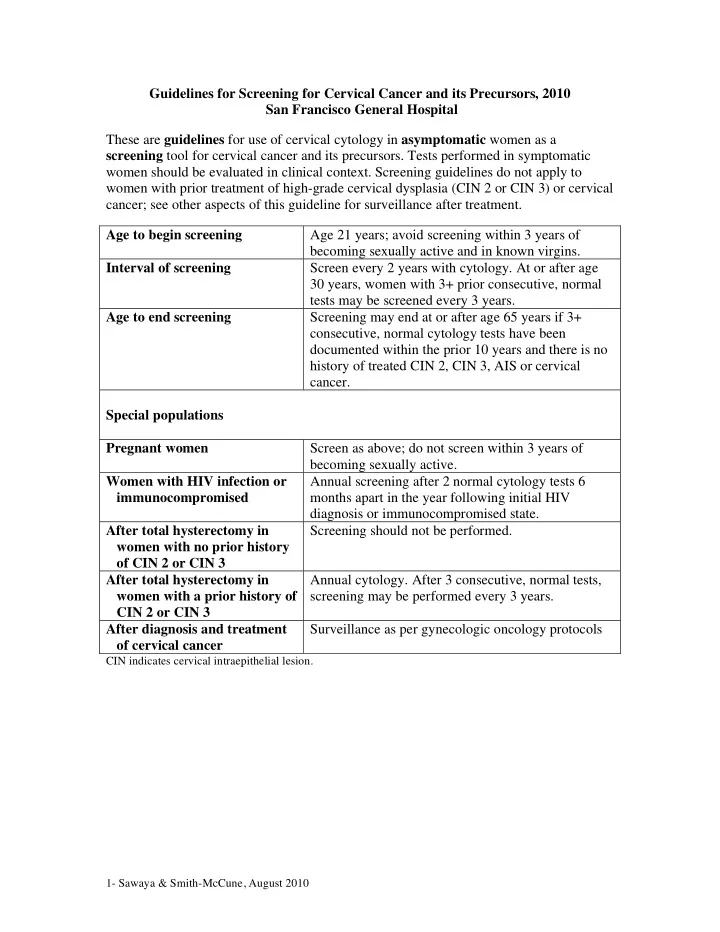
Guidelines for Screening for Cervical Cancer and its Precursors, 2010 San Francisco General Hospital These are guidelines for use of cervical cytology in asymptomatic women as a screening tool for cervical cancer and its precursors. Tests performed in symptomatic women should be evaluated in clinical context. Screening guidelines do not apply to women with prior treatment of high-grade cervical dysplasia (CIN 2 or CIN 3) or cervical cancer; see other aspects of this guideline for surveillance after treatment. Age to begin screening Age 21 years; avoid screening within 3 years of becoming sexually active and in known virgins. Interval of screening Screen every 2 years with cytology. At or after age 30 years, women with 3+ prior consecutive, normal tests may be screened every 3 years. Age to end screening Screening may end at or after age 65 years if 3+ consecutive, normal cytology tests have been documented within the prior 10 years and there is no history of treated CIN 2, CIN 3, AIS or cervical cancer. Special populations Pregnant women Screen as above; do not screen within 3 years of becoming sexually active. Women with HIV infection or immunocompromised Annual screening after 2 normal cytology tests 6 months apart in the year following initial HIV diagnosis or immunocompromised state. After total hysterectomy in women with no prior history
- f CIN 2 or CIN 3
Screening should not be performed. After total hysterectomy in women with a prior history of CIN 2 or CIN 3 Annual cytology. After 3 consecutive, normal tests, screening may be performed every 3 years. After diagnosis and treatment
- f cervical cancer