Disclosures This is How I Do It Bard Peripheral Vascular - - - PowerPoint PPT Presentation

disclosures
SMART_READER_LITE
LIVE PREVIEW

Disclosures This is How I Do It Bard Peripheral Vascular - - - PowerPoint PPT Presentation

4/18/2015 Disclosures This is How I Do It Bard Peripheral Vascular - Research, Consultant Rertrograde Tibio-Pedal Cardiovascular Systems, Inc. - Research, Consultant Access Cook Medical - Research, Consulting Covidien


slide-1
SLIDE 1

4/18/2015 1

This is How I Do It Rertrograde Tibio-Pedal Access

Fadi Saab MD, FACC, FASE, FSCAI Clinical Assistant Professor- Michigan State University School of Medicine Department of Internal Medicine- Metro Heart and Vascular Metro Health Hospital

Disclosures

  • Bard Peripheral Vascular - Research, Consultant
  • Cardiovascular Systems, Inc. - Research, Consultant
  • Cook Medical - Research, Consulting
  • Covidien – Consulting
  • Terumo – Consulting
  • Spectranetics – Research, Consulting

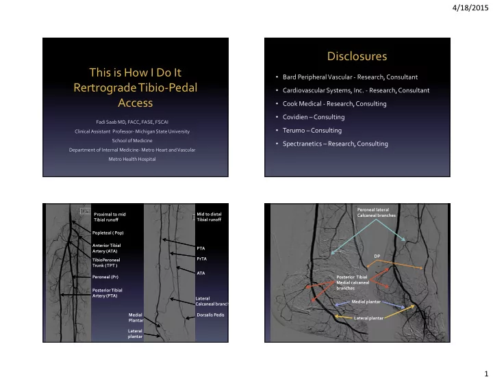
PTA PrTA ATA Mid to distal Tibial runoff Proximal to mid Tibial runoff Dorsalis Pedis Medial Plantar Lateral plantar Popleteal ( Pop) Anterior Tibial Artery (ATA) TibioPeroneal Trunk ( TPT ) Peroneal (Pr) Posterior Tibial Artery (PTA) Lateral Calcaneal branch Peroneal lateral Calcaneal branches Posterior Tibial Medial calcaneal branches DP Medial plantar Lateral plantar

slide-2
SLIDE 2

4/18/2015 2

AT Tibioperoneal trunk PT

PERONEAL AT TPT Pr PT TPT Vein RLE A=AT B=TPT C=PT D=Pr A B C D

Tibial Vessels Evaluation and Access

  • Leg Orientation
  • Probe Selection
  • Utilization of US in delivering therapy
slide-3
SLIDE 3

4/18/2015 3

Technique

  • Assessing the ideal spot for

retrograde tibiopedal arterial access site is mainly done by ultrasound.

  • The operator can evaluate the

vessels with color and pulse-wave Doppler in multiple planes.

  • This is of paramount importance as

it decreases the likelihood of venous puncture, venous sheath placement, AV fistulas, and tibial artery spasm. AMP Group, 2012

Technique

  • Arterial spasm decreases the

likelihood of success, especially when the vessel lumen is already compromised.

  • At our institution we use the

Philips linear 15i7 MHz hockey stick probe and the Philips iU22 X-Matrix (Philips Electronics, Andover, MA).

AMP Group, 2012

Technique

  • As we move the probe cranially, it is easy to

visualize how the tibial veins start to separate from the tibial arteries, allowing easier cannulation of the tibial vessels in a spot where the veins are not located in the planned needle trajectory.

  • While moving cranially, it is essential to keep in

mind the four major anatomical compartments below the knee.

  • These compartments lay within the

gastrocnemius muscle and most of the time end at the insertion points of the distal gastrocnemius heads.

Technique

  • It is imperative to avoid accessing beyond the gastrocnemius

heads in order to decrease the likelihood of a complication resulting in compartment syndrome, which in turn can lead, to emergent surgical intervention and in rare occasions even amputation.

  • Arterial access below the gastrocnemius heads, allows the
  • perator to have complete control to address potential bleeding

complications during and after tibial access procedures.

  • A vascular technologist is present during the access process.
slide-4
SLIDE 4

4/18/2015 4

Technique

  • The short and long access views of these

vessels will reveal the access point.

  • The operator will monitor the introduction
  • f the access needle .
  • Retrograde tibial access will identify a

hibernating lumen of these vessels not

  • therwise identified with traditional

angiography due to proximal vessel

  • cclusion.
  • Tibial lesions also can be distal and easy to

identify on US evaluation.

Technique

  • It is our practice to visualize the wire under US

guidance while traveling inside the vessel.

  • Once access is gained into the tibial vessel, the

micro sheath is introduced into the vessel.

Angiographic Confirmation

  • We then inject contrast to confirm our

intraluminal position.

  • If the patient blood pressure allows,

we inject 300-400 micrograms of nitroglycerin into the tibial vessel.

  • Depending on the operator, usually a

4 French micro sheath will be inserted into the tibial vessel.

Saab et al

Anterior Tibial Artery Access

  • The tibial vessels are accessed

in the following fashion:

  • Typically the foot is prepped

and draped separately.

  • The orientation of the foot is

adjusted depending on the target tibial vessel.

Saab et al

slide-5
SLIDE 5

4/18/2015 5

Anterior Tibial Artery Access

  • In cases of the dorsalis pedis (DP)
  • r the distal anterior tibial artery

(AT), the foot is maintained in natural orientation with the heel of the foot on the mattress with slight dorsiflexion Saab et al

Vessel Interrogation

Saab et al

Posterior/Peroneal Tibial Artery Access

  • To access the posterior tibial

artery (PT) the foot is rotated laterally and the leg will be bent slightly at the knee level for patient comfort.

  • To access the peroneal artery the

foot needs to be rotated laterally further to separate the fibula and

  • tibia. This maneuver will facilitate

direct cannulation of the artery.

Saab et al

Vessel Interrogation

Saab et al

slide-6
SLIDE 6

4/18/2015 6

The Operater will choose a lower frequency Probe to image the tibial vessels as they dive into the major Compartments

Saab et al

Ultrasound Guided Tibial- Pedal access procedure

J.A.Mustapha, MD J.A.Mustapha, MD

slide-7
SLIDE 7

4/18/2015 7

The science of tibial access

45 45 60-70 Degrees

J.A.Mustapha, MD

A B

J.A.Mustapha, MD J.A.Mustapha, MD J.A.Mustapha, MD

slide-8
SLIDE 8

4/18/2015 8

J.A.Mustapha, MD J.A.Mustapha, MD

Needles

Saab et al Saab et al

slide-9
SLIDE 9

4/18/2015 9

Saab et al 3 O’clock 9 O’clock 12 1 2 11 10 Figure X: the best ultrasound guided entry points into the tibial artery

  • 2 and 10 O’clock are the 3rd best entry points into the tibial artery
  • 12 O’clock is the best entry point into the tibial artery
  • 1 and 11 O’clock are the 2nd best entry points into the tibial artery

Saab et al

slide-10
SLIDE 10

4/18/2015 10

Extra Vascular Ultrasound EVUS

  • The process of using US to obtain, guide and

deliver therapy

Saab et al

slide-11
SLIDE 11

4/18/2015 11

Wires

Reverberation artifacts appear as multiple equally spaced lines Saab et al

Why is this better than contrast??

Saab et al Saab et al

No Contrast Yet

Saab et al

slide-12
SLIDE 12

4/18/2015 12

Catheters

Saab et al

Catheters

Saab et al A B C D Popliteal cross sectional view Popliteal longitudinal view

J.A.Mustapha MD

Longitudinal view of the “white stop sign” Short access view of the “white stop sign”

White Stop Sign

J.A.Mustapha MD

slide-13
SLIDE 13

4/18/2015 13

Our Experience

  • The Tibio-Pedal Arterial Minimally Invasive

Retrograde Revascularization Technique (TAMI Technique) has been established at our institution since 2011

  • Extra vascular ultrasound in the cath lab (EVUS)

has been established at our institution since 2011

Flouro Time with TAMI and EVUS

5 10 15 20 25 30 35 2012 2013 33.09 27.4 22.3 15.43 PV TAMI Time in minutes Saab et al

Thank You Fadi.saab@metrogr.org 313-590-5902