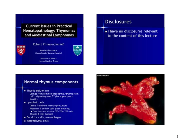

Disclosures Current Issues in Practical Hematopathology: Thymomas � I have no disclosures relevant and Mediastinal Lymphomas to the content of this lecture Robert P Hasserjian MD Associate Pathologist Massachusetts General Hospital Associate Professor Harvard Medical School Normal thymus Normal thymus components � Thymic epithelium – Derives from common endodermal ‘thymic stem cell’ originating from 3 rd pharyngeal pouch – Keratin+ � Lymphoid cells – Derive from bone marrow precursors – Precursor T and NK cells (vast majority) � Enter thymus as primitive CD3-/CD4-/CD8- cells – Thymic B cells (sparse) � Dendritic cells, macrophages � Mesenchymal cells 1
Normal thymus Normal thymus Medulla CD4+ or CD8+ CD1a-, TdT-, CD99- Low Ki67 PI Low rate of apoptosis Cortex CD4+CD8+ CD1a+, TdT+, CD99+ High Ki67 PI High rate of apoptosis Cytokeratin Normal thymus Role of pathologist in Cortex & medulla Cortex thymoma management CD3 CD4 TdT � Identify tumor as thymoma – Tumor arising from thymic epithelium � Determine presence and degree of invasion (stage) � Determine histologic subtype CD5 CD8 CD1a – WHO Working Group Classification 2004, as modified from original classification of 1999 2
Thymoma Thymoma staging: a collaboration Types of invasion* between radiologists, surgeons, and pathologists Stage T3 Invasion of innominate vein, � Fully encapsulated (T1, Masaoka I) indistinct margins Stage T2 � Minimally invasive (T2, Masaoka II) – Penetrates capsule to invade pericapsular connective tissue, fat, or adjacent thymus � Invades other organs (lung, pleura, pericardium, diaphragm, chest wall) Invasion of adjacent normal (T3, Masaoka III) thymus *TNM Classification and Modified Masaoka Staging systems 3
WHO Thymoma Classification Thymoma: Stage and Type do make a difference. . . WHO Features Prognosis Type A Spindle cell morphology, Good medullary-type lymphocytes Type B1 Recapitulation of normal thymic Good architecture (‘organoid’) Type B2 Epithelial cells frequent, Fair cortical-type lymphocytes Type B3 Atypical epithelial cells Fair to predominate poor Type AB Mixed features (usually A + B1) Good ‘Type C’ Carcinoma Poor Hasserjian R, Strobel P, Marx A. Semin Thor Cardiovasc Surg 2005; 17: 2-11 Thymoma Type A Thymoma Type A � Well-circumscribed – Mean size 10 cm – 80% T1 � Derive from medullary- type epithelial cells � Lymphocyte-poor – Lymphocytes mostly have phenotype of medullary thymocytes (TdT-, CD1a-) � Excellent prognosis, ‘benign thymoma’ 4
Thymoma Type A Thymoma Type B1 � Well-circumscribed – 60% T1, 25% T2 � Derive from ‘common’ thymic epithelial cell � Lymphocyte-rich Key features of Type A: – Mainly cortical-type, � Bland-appearing ovoid to spindled cells with more mature T � Absence of mitotic figures cells in medullary areas � Sparse lymphocytes in most areas � Excellent prognosis � Epithelial cells may express CD20 – Behavior depends on stage and resectability Cytokeratin Type B1 Thymoma Type B1 5
Medullary differentiation in B1 Normal thymus Medullary differentiation in B1: TdT M M M M M Thymoma Type B1: area of medullary (‘organoid’) differentiation Thymoma Type AB Key features of Type B1: � Bland-appearing round epithelial cells lost in a sea of cortical thymocytes � Mitotic figures usually frequent in thymocytes � Epithelial cells may have small nucleoli � Scattered pale spots that recapitulate normal thymic medulla 6
Thymoma Type AB Features of Thymoma Type B2 � Circumscribed or invasive – T1, T2, T3 about equal Key features of Type AB: – 10% disseminated � Bland-appearing epithelial cells range � Derive from cortical- from round to spindled in discrete areas type epithelial cell � Lymphocyte-rich areas usually resemble � Lymphocytes present, B1, not B2 thymomas but fewer than B1 � Epithelial cells may express CD20, like – Immature cortical type Type A thymomas � Intermediate prognosis Thymoma Type B2 Thymoma Type B2 7
Thymoma Type B2 Thymoma Type B2 TdT Key features of Type B2: � Epithelial cells more frequent, but many cortical thymocytes are still present � Tumor cells may have nucleoli and form clusters � Lack areas of medullary differentiation Thymoma Type B3 Features of Thymoma Type B3 � Infiltrative – T3 and T2 common – 15% disseminated � Derive from cortical- type epithelial cell � Lymphocyte-poor � Intermediate prognosis – Often unresectable – Local recurrence frequent 8
Thymoma Type B3 Thymoma Type B3 TdT Key features of Type B3: � Epithelial cells form sheets � Lymphocytes are sparse � Epithelial cells are atypical, with ‘raisinoid’, crinkled, or (less frequently) vesicular nuclei and often distinct cell borders Thymoma Type A Thymoma Type B3 Spindled cell proliferations in the anterior mediastinum � Type A thymoma – Densely cellular, but cytologically bland – No or very rare mitotic figures – May express CD20 � Type B3 thymoma – Cells oval, not spindled – Nuclear atypia and usually increased mitotic activity � Thymic sarcomatoid carcinoma – Overtly malignant cytology, high mitotic rate � Solitary fibrous tumor – Keratin-, CD34+, CD99+ Weissferdt A Appl Imm Mol Morphol 2011;19:329 9
Thymic squamous cell carcinoma Thymic carcinoma � Squamous cell � Basaloid � Mucoepidermoid � Lymphoepithelioma-like � Sarcomatoid/carcinosarcoma � Clear cell � Adenocarcinoma – Papillary adenocarcinoma � Neuroendocrine carcinomas – Poorly-differentiated (small cell or large cell types) – Well-differentiated (thymic carcinoid) � Carcinoma with t(15;19) Thymic squamous cell carcinoma Thymic lymphoepithelial carcinoma 10
Thymoma versus Thymic carcinoma versus thymic carcinoma other tumors � The thymus can be a site of invasion of ‘Benign’ thymoma Thymic carcinoma metastasis from lung or other carcinomas � Clinical and radiologic features are critical in � Cytologically bland � Cytologically malignant this distinction epithelial cells epithelial cells – No specific keratin expression profile � Recapitulate thymic � Do not resemble normal – May express calretinin (1/3 of thymic carcinomas) architecture thymus – TTF1 negative � Typically CD5- and � Often CD5+ and/or CD117- CD117+ � β 5t specific for thymic origin, but only � β 5t positive � β 5t negative expressed in B2/B3, not thymic carcinomas � Autoimmune � No autoimmune � CD5 positivity favors thymic origin associations common associations – 40-50% CD5 positive, negative in lung and other carcinomas Pan CC Hum Pathol 2003; 34:1155; Yamada Y AJSP 2011;35:1296 Yamada Y Am J Surg Pathol 2011;35:1296 A B1 Increasing epithelial cell density Thymic lymphomas Decreasing lymphocyte number � Relatively common B2 – Mediastinal large B-cell lymphoma (MLBCL) – Classical Hodgkin lymphoma (CHL) Epithelial atypia – T-lymphoblastic lymphoma (T-LBL) Thymic carcinoma � Uncommon B3 – Thymic MALT lymphoma – Anaplastic large cell lymphoma and other peripheral T/NK lymphomas – ‘Grey zone’ lymphoma (DLBCL/CHL overlap) 11
Mediastinal mass from 42 year-old woman T-lymphoblastic lymphoma � Precursor T-cell neoplasm – CD3+, TdT+, CD1a+, CD99+, CD4/CD8 variable, keratin- � Children and young adults (M>F) � May have associated leukemic involvement with circulating T- lymphoblasts � Treatment is chemotherapy, not surgery Mediastinal mass: TdT stain Pifalls in distinguishing T-LBL from thymoma � Thymomas may occur in children and young adults � Reactive T-lymphocytosis can be associated with thymomas � Immunophenotype and morphology of T-LBL and lymphocytes of cortical thymomas can be identical T-LBL Thymoma � Infiltrative � Lobulated � No or few keratin+ cells � Keratin+ tumor cells � Clonal TCR � Polyclonal T cells rearrangements 12
T-LBL T-LBL B1 Thymoma T-LBL TdT TdT B1 Thymoma Mediastinal large B cell lymphoma (MLBCL) � Tumor of young adults (F > M) � Locally aggressive with dissemination to extranodal sites – Often very bulky mass, SVC syndrome Cytokeratin Cytokeratin � Thought to derive from thymic B-cell – Gene expression profiling has shown that MLBCL is distinct from DLBCL and has some similarities to classical Hodgkin lymphoma � Immunophenotype – CD19+, PAX5+, CD20+, CD79a+, CD30 often weak+ – Ig clonally rearranged, but not expressed 13
Mediastinal large B-cell lymphoma Mediastinal large B-cell lymphoma CD45 CD20 CD30 CD20 TdT Ki67 Courtesy of Dr Aliyah Sohani 14
Nodular sclerosis classical Hodgkin lymphoma The molecular signature of MLBCL differs from that of other DLBCL Nodal DLBCL MLBCL Over-expressed MAL, FIG-1 Fibronectin Collagens c-REL, TRAF1, STAT1 Under-expressed IgM MHC class II Savage K et al. Blood 2003; 102:3871 Courtesy of Dr Laurence de Leval, Lausanne, Switzerland Rodig SJ et al. AJSP 2007; 31:106 Nodular sclerosis classical Hodgkin lymphoma Classical Hodgkin lymphoma MLBCL 15
Recommend
More recommend