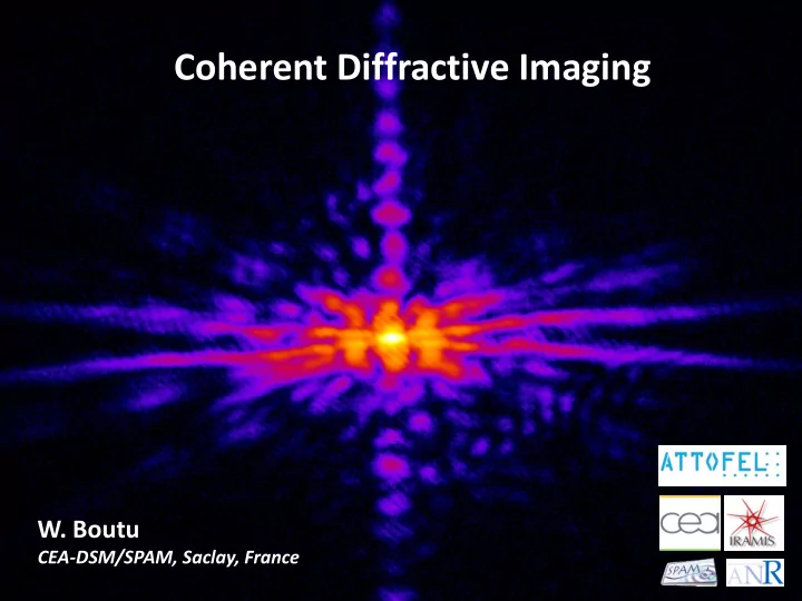

Coherent Diffractive Imaging W. Boutu CEA-DSM/SPAM, Saclay, France
OUTLINE Introduction Fundamentals of lensless imaging • Far field imaging • The phase problem • Resolution and limitations Solving the phase problem with iterative algorithms • Basics on phase retrieval iterative algorithms • Some examples Holographic techniques • Fourier Transform Holography • Holography with Extended References
OUTLINE Introduction Fundamentals of lensless imaging • Far field imaging • The phase problem • Resolution and limitations Solving the phase problem with iterative algorithms • Basics on phase retrieval iterative algorithms • Some examples Holographic techniques • Fourier Transform Holography • Holography with Extended References
WHY IMAGING WITH X-RAYS Spatial resolution d t : Rayleigh criterion: the central peak of the Airy pattern from one circular aperture in located at the first minimum ring of the Airy pattern from a second hole at a distance d t . d d t Z 𝑨𝑓𝑠𝑝 = 1.22𝜇𝑨 First zero at: 𝑠 = δ 𝑢 • 𝑒 𝜀 𝑢 = 0.61𝝁 𝑂. 𝐵. • Numerical aperture: 𝑂. 𝐵. = 𝑜 sin 𝜄 ~𝑒/2𝑨
WHY IMAGING WITH X-RAYS Spatial resolution d t : High penetration depth: In the Xray domain, n very close to 1. 𝑜 𝜕 = 1 − 𝜀 + 𝑗𝛾 Usually written: y x z Near field exit wave after a sample: Ψ 𝑨, 𝑢 = Ψ 0 𝑓 −𝑗(𝜕𝑢−𝑨/𝑑) 𝑓 𝑗(2𝜌 𝜇 )𝜀𝑨 𝑓 −(2𝜌 Plane wave: Ψ 𝒔, 𝑢 = Ψ 0 𝑓 −𝑗(𝜕𝑢−𝒍.𝒔) 𝜇 )𝛾𝑨 phase shift absorption Attenuation coefficient: Beer Lambert law: 𝐽 𝑨 = 𝐽 0 𝑓 −µ𝑨 Absorption coeff. very small with X-rays Water window (Kirz et al. , Quaterly Reviews of Biophysics (1995))
IMAGING WITH OPTICS Lens equivalent in the VUV domains: Metallic optics at grazing incidence Reflective optics with multilayer coating Lens equivalent in the X-ray domains: Kirkpatrick-Baez mirror Fresnel zone plate (Chao et al. , Nature (2005)) Image of a 15.1nm half period test object with 2 zone plates at l =1.52 nm. ZP outermost ZP outermost Spatial resolution ≈ 12 nm. width=25nm width=15nm
FRESNEL ZONE PLATES Resolutions: 𝜇 with d r n outermost zone width 𝑂. 𝐵. = Numerical aperture: 2𝜀𝑠 𝑜 𝜀 𝑢 = 1.22𝜀𝑠 𝑜 Transverse resolution: 2 𝜀 𝑚 = 4.88 𝜀𝑠 𝑜 Depth of focus: 𝜇 Resolution dependent on the ZP quality. Limitations: Currently, the best ZP have a d r n of 12nm, but a efficiency of less than 1%. Usual ZP: d r n ≈ 30nm, efficiency ≈ 10% Low photon flux, need for long accumulation times. Pb for imaging biological samples.
OUTLINE Introduction Fundamentals of lensless imaging • Far field imaging • The phase problem • Resolution and limitations Solving the phase problem with iterative algorithms • Basics on phase retrieval iterative algorithms • Some examples Holographic techniques • Fourier Transform Holography • Holography with Extended References
LENSLESS IMAGING r ⊥ R ⊥ r 𝑨 0 𝑎 In free space, the scalar propagation equation can be written as the Helmoltz equation: 𝛼 2 𝝎 𝒔 + 𝑙 2 𝝎 𝒔 = 0 Taking the Fourier transform in the transverse plan only, one gets: 2 + 𝜖 𝑨 2 + 𝑙 2 𝜔 𝒓 ⊥ ; 𝑨 = 0 −𝒓 ⊥ 𝒓 ⊥ ; 𝑨 = 𝜔 𝒓 ⊥ ; 0 𝑓 𝑗𝜆𝑨 with 𝜆 = 𝑙 2 − 𝒓 ⊥ 𝜔 2 The general solution, in the propagation direction, is: Taking the wave at the exit of the sample, 𝜔 0 , for the boundary conditions: 𝜔 𝒔 ⊥ ; 𝑨 = ℱ −1 𝜔 0 𝒓 ⊥ 𝑓 𝑗𝜆𝑨
LENSLESS IMAGING 𝜔 𝒔 ⊥ ; 𝑨 = ℱ −1 𝜔 0 𝒓 ⊥ 𝑓 𝑗𝜆𝑨 𝑙 2 − 𝒓 ⊥ 2 with 𝜆 = Paraxial approximation : expand k to the first non zero order in 𝒓 ⊥ . 2 𝒓 ⊥ 𝑓𝑦𝑞 𝑗𝑙𝑨 1 − 𝒓 ⊥ 𝜔 𝒔 ⊥ ; 𝑨 = ℱ −1 𝜔 0 2𝑙 2 1 𝜔 0 𝒔 ⊥ ∗ 𝑔 𝒔 ⊥ ; 0 with f the Fresnel propagator. 𝜔 𝒔 ⊥ ; 𝑨 = 2𝜌 𝜔 𝒔 ⊥ ; 𝑨 = 𝑒𝑺 ⊥ 𝜔 0 𝑺 ⊥ ; 0 𝑓 𝑗𝑙 𝒔 ⊥ −𝑺 ⊥ 2 which can be written as 2𝑨 with 𝑺 ⊥ the variable in the sample plane (omitting some phase terms): 2 𝜔 𝒔 ⊥ ; 𝑨 = ℱ 𝜔 0 𝑺 ⊥ 𝑓𝑦𝑞 𝑗 𝑙𝑺 ⊥ or 2𝑨 𝒓 ⊥ =𝑙𝒔 ⊥ /𝒜 Fraunhofer approximation : the phase modulation due to the exp. term is small. 𝑏 2 i.e. the Fresnel number 𝑜 = 𝜇𝑨 ≪ 1 ( a being the typical size of the sample). 𝑏 2 𝜔 𝒔 ⊥ ; 𝑨 = ℱ 𝜔 0 𝑺 ⊥ 𝜇 ) , the diffraction field reads: So in the far field ( 𝑨 ≫
THE PHASE PROBLEM 𝜔 𝒔 ⊥ ; 𝑨 = ℱ 𝜔 0 𝑺 ⊥ The diffracted wave by a sample is, in the far field, the Fourier transform of the sample transmittance. Measured diffracted intensity 2 ℱ 𝜔 0 𝑺 ⊥ Can the phase be recovered? Phase lost 𝜔 0 𝑺 ⊥ ( k ) sample
RESOLUTION Δ𝑠 Maximum transverse resolution: sample q max 𝜎 𝑢 = 𝜇𝑎 𝑜 𝐺 Δ𝑠 𝑜 𝐺 Δ𝑠 Z 0 Longitudinal resolution: CCD 𝜎 𝑚 = 2𝜇𝑎 2 𝑜 𝐺 Δ𝑠 2 Example with typical values for experiment in our lab: l = 32 nm Z = 2 cm Maximum resolution : 47 nm D r = 13.5 µm n F = 1000 • Limitations: Missing central spot due to saturation • SNR and dose requirements • Coherence
MISSING CENTRAL SPOT Due to the brightness of most Xray sources and the low scattering efficiency, the direct beam saturates the CCD use of a beam block to stop it. The low frequency data are missing! ℱ −1 ℱ
MISSING CENTRAL SPOT Ways around: Create the final diffraction pattern by summing different acquisition times : (Chen et al. , PRA (2009)) This image is the addition of 1000 frames with 1.2 s exposure each and 30 frames with 78s exposure, for a total of 59mn. Low pass filtering: Use of unconstrained modes: find the least constrained modes and subtract them from the reconstruction (Thibault, PhD dissertation)
SIGNAL TO NOISE resolution 56 nm 56 nm In the diffraction pattern, signal up to 8.9 µm -1 , corresponding to a resolution of 56nm. But the actual resolution is only 78nm. Resolution limited by SNR Rose criterion : a SNR of at least 5 is needed. 8.9 µm -1 -8.9 µm -1 0 Sources of noise: Ways around: • • Diffuse light, coming e.g. from the focusing optics Improve your setup • • Hardware binning Beam properties fluctuations Photon noise, scales as 𝑜𝑣𝑛𝑐𝑓𝑠 𝑝𝑔 𝑞𝑖𝑝𝑢𝑝𝑜𝑡 • • Increase the photon flux • • Readout noise from the CCD Post experiment image processing • Increase the statistic, and correlate the different images
DOSE Increasing the number of photons => increasing the intensity of the radiation received. But samples, especially biological samples, are sensitive to radiations. = 𝜈𝑂 0 𝐹 µ = absorption coeff. absorbed energy Dose (in Gray) = 𝜍𝐵 N 0 = number of photons per unit area A mass E = photon energy ≈5nm = maximum resolution for CDI? Destruction of crystalline order in protein crystals Visible structural changes in biological specimens (Shen et al. , J. Synchrotron Rad. (2004))
DOSE Increasing the number of photons => increasing the intensity of the radiation received. But samples, especially biological samples, are sensitive to radiations. = 𝜈𝑂 0 𝐹 µ = absorption coeff. absorbed energy Dose (in Gray) = 𝜍𝐵 N 0 = number of photons per unit area A mass E = photon energy ≈5nm = maximum resolution for CDI? Use XFEL : the pulse duration is so short that the diffraction pattern is taken before the damages are made! Single FLASH exposure (Shen et al. , J. Synchrotron Rad. (2004)) Chapman et al. , Nature Physics (2006)
TEMPORAL COHERENCE Coherence requirement: Using the small angle approximation 𝜇 2 and the resolution definition, one gets: Coherence length ≈ 2Δ𝜇 𝜇 2 Δ𝜇 ≥ 𝑏 𝜇 2Δ𝜇 ≥ 𝑏 sin 𝜄 𝜎 𝑢 a/2 𝑏/2 sin 𝜄 There are some ways around this limitation: For instance, Chen et al. , PRA (2009) They assume that the diffraction patterns from the different order do not interfere. SEM image reconstruction Input Harmonics spectrum
SPATIAL COHERENCE Some XUV sources are almost fully spatially coherent, like HHG, but not synchrotrons. Solution: use a slit, because the illumination within the half radius of the Airy pattern can be considered as coherent, according to the van Cittert-Zernike theorem. 𝑚 𝑢 = 0,61𝜇𝐸 with D = distance hole-sample 𝜚 ℎ𝑝𝑚𝑓 One way around this limitation: consider a multimodal field with mutually uncoherent modes (Whitehead et al. , PRL (2009)) high coherence low coherence
CONCLUSION OF PART I In the far field, the diffractive field is the Fourier transform of the wave after the sample. 𝜔 𝒔 ⊥ ; 𝑨 = ℱ 𝜔 0 𝑺 ⊥ In a measurement, the spatial phase is lost. Be careful not to damage your sample with radiation… or explode it on purpose!
OUTLINE Introduction Fundamentals of lensless imaging • Far field imaging • The phase problem • Resolution and limitations Solving the phase problem with iterative algorithms • Basics on phase retrieval iterative algorithms • Some examples Holographic techniques • Fourier Transform Holography • Holography with Extended References
Recommend
More recommend