
2/14/2014 1
Clinical Pathological Conference
Cheryl Jay, MD, Professor of Neurology, UCSF Director of Neurology Clinics, San Francisco General Hospital Andrew W. Bollen, MD, DVM Professor of Pathology, UCSF Mark Burish MD, PhD Neurology Resident, UCSF
- 55yo male with a history of cryptogenic
strokes at age 45
- Chief complaint: left leg swelling, progressive
multifocal numbness and weakness
- Jan 2012: progressive left leg swelling and erythema starting
in the calf and extending to the foot, initially intermittent then became constant and slightly painful
- March 2012: admitted to an outside hospital for leg swelling
and pain.
– Lower extremity Doppler U/S negative – Noted to have leukopenia with ANC of 1000 – Muscle biopsy with “focal mild endomysial inflammation in 1 muscle fascicle with histiocytes” – Treated for possible vasculitis with prednisone, mild improvement in symptoms
- May 15 2012: Feeling of being off balance, needing to hold
- n to objects while walking progressing over 1 week to left
foot drop
- May 29 2012: Numbness on right medial palm and ring and
pinky fingers
- June 5 2012: softening of his voice
- June 8 2012: Admitted to UCSF
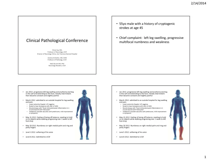