Cervical Spine Cervical Spine C1 - C7 Atlas and Axis Ligamentous - - PowerPoint PPT Presentation

cervical spine
SMART_READER_LITE
LIVE PREVIEW

Cervical Spine Cervical Spine C1 - C7 Atlas and Axis Ligamentous - - PowerPoint PPT Presentation

Cervical Spine Cervical Spine C1 - C7 Atlas and Axis Ligamentous Anatomy Anterior longitudinal ligament Reinforces anterior discs, limits extension Posterior longitudinal ligament Reinforces posterior discs, limits flexion


slide-1
SLIDE 1

Cervical Spine

slide-2
SLIDE 2

Cervical Spine

  • C1 - C7
slide-3
SLIDE 3

Atlas and Axis

slide-4
SLIDE 4

Ligamentous Anatomy

  • Anterior longitudinal ligament

– Reinforces anterior discs, limits extension

  • Posterior longitudinal ligament

– Reinforces posterior discs, limits flexion

  • Ligamentum nuchae = supraspinous ligament

– Thicker than in thoracic/lumbar regions – Limits flexion

  • Interspinous/intertransverse ligaments

– Limit flexion and rotation/limits lateral flexion

  • Ligamentum flavum

– Attach lamina of one vertebrae to another, reinforces articular facets – Limits flexion and rotation

slide-5
SLIDE 5

Ligamentous Anatomy

  • a = ligamentum

flavum

  • b = interspinous

ligaments

  • c = supraspinous

ligament

slide-6
SLIDE 6
  • Palpable C7
  • Anterior Curvature

– Shock absorption

  • Ligaments

– Ligamentum Nuchae – “Whiplash”

  • Vertebral Arteries
slide-7
SLIDE 7
  • Spinal Nerves

– C1-T1 – Cervical Plexus

  • C1-C4
  • C4 -Phrenic Nerve - Breathing

– Brachial Plexus

  • C5-T1

C3

slide-8
SLIDE 8

Dermatomes Myotomes

C5 – Abduction C6 – Elbow Flexion/Wrist Extension C7 – Elbow Extension/Wrist Flexion C8 – Finger Flexion T1 – Finger Abduction C1-2 – Neck Flexion C3 – Lateral Neck Flexion C4 – Shoulder Elevation C1 – top of head C2 – Temporal C3 – Side of jaw/neck C4 – top of shoulders

slide-9
SLIDE 9

Brachial Plexus

slide-10
SLIDE 10

Brachial Plexus

ROOTS TRUNKS DIVISIONS CORDS BRANCHES C5 C6 C7 C8 T1

Upper Middle Lower

Anterior Posterior Anterior Posterior Anterior Posterior

Lateral Posterior Medial

Suprascapular Lateral Pectoral Musculotaneous Axillary Radial Median Ulnar

Long Thoracic Medial Pectoral Medial Antebrachial Medial Brachial Cutaneous Thoracodorsal Subscapular

Dorsal Scapular

slide-11
SLIDE 11
slide-12
SLIDE 12
  • Muscles

Trapezius Sternocleidomastoid Scalenes Splenius Semispinalis, Spinalis, Longissimus

slide-13
SLIDE 13

Cervical Injuries

  • Fairly uncommon in athletics(6-7%) - but greater than

90% of all fatalities are cervical related.

  • Cervical injuries are primarily technique related:

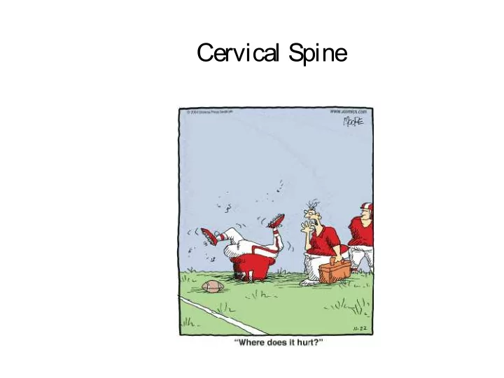
– Spearing – Tackling or falling head first.

  • Must have an emergency plan:

– All personnel know roles and equipment use. – All unconscious athletes - suspect head/neck – Always suspect the worse until proven otherwise

slide-14
SLIDE 14

Cervical Injuries

  • Common MOIs

– Axial Loading – Flexion Force – Hyperextension Force – Flexion-Rotation Force – Hyperextension-Rotation – Lateral Flexion

slide-15
SLIDE 15
slide-16
SLIDE 16

C-Spine Injuries

  • Cervical Fracture or Dislocation

– Weakness or Paralysis

  • Cervical Nerve Root Injury

– Herniated Disc – Laceration – Cord Shock (Central Cord Syndrome) – Hemorrhage – Contusion – Cervical Stenosis

slide-17
SLIDE 17
slide-18
SLIDE 18
slide-19
SLIDE 19

C-Spine/Neck Injuries

  • Cervical Strain

– Active motion most painful

  • Cervical Sprain (Whiplash)

– Passive and active motion painful

  • Torticollis (WryNeck)

– Muscle spasm and facet irritation

  • Brachial Plexus Stretch or Compression
  • Contusions to Throat
slide-20
SLIDE 20

Evaluation Techniques

  • HOPS

– History, Observation, Palpation, Special Tests

  • Your first priority!

– Establish the integrity of the spinal cord and nerve roots – History and several specific tests provide information

slide-21
SLIDE 21

History

slide-22
SLIDE 22

History

  • Location of pain
  • Onset of pain
  • Mechanism of injury (etiology)
  • Consistency of pain
  • Prior history of cervical spine injury
slide-23
SLIDE 23

Location of Pain

  • Localized pain

– Typically indicative of muscular strain, ligamentous sprain, facet joint injury, fracture and/or subluxation or dislocation

  • Radiating pain

– Heightened risk of likely spinal cord, cervical nerve root and/or brachial plexus injury

slide-24
SLIDE 24

Onset of Pain/Mechanism of Injury

  • Acute onset

– Generally associated with one specific mechanism of injury/event

  • Chronic or insiduous (unknown) onset

– Generally related to overuse injuries (accumulative microtrauma) and/or postural abnormalities and deficiencies

slide-25
SLIDE 25

Consistency of Pain

  • Pain from inflammation (strain, sprain,

contusion) generally persists despite changes in cervical spine position

  • Pain of mechanical nature (nerve root

compression) varies depending upon cervical spine positioning and can be minimized or eliminated

slide-26
SLIDE 26

Prior History of Cervical Spine Injury

  • Must evaluate for residual symptoms

associated with previous injury

  • Must appreciate structural changes (scar

tissue, etc.) which may predispose individual to current injury and symptoms

slide-27
SLIDE 27

Inspection

slide-28
SLIDE 28

Inspection

  • Cervical spine curvature
  • Position of head relative to shoulders
  • Soft tissue symmetry
  • Level of shoulders
slide-29
SLIDE 29

Cervical Spine Curvature

  • Normal cervical spine has lordotic curve
  • Increased lordotic curve (forward head)

indicative of poor posture and muscular weakness or imbalance

  • Lessened lordotic curve indicative of

muscular spasm/guarding and/or nerve root impingement

slide-30
SLIDE 30

Lordotic Curve

slide-31
SLIDE 31

Position of Head Relative to Shoulders

  • Head should be seated symmetrically on

cervical spine

  • Lateral flexion from unilateral spasm of

muscles – strain and/or spasm (guarding)

  • Rotation from unilateral spasm of

sternomastoid muscle – strain and/or spasm (guarding) or torticollis

slide-32
SLIDE 32

Torticollis

slide-33
SLIDE 33

Soft Tissue Symmetry

  • Observe for bilaterally comparable muscle

mass, tone and contour

– Dominant extremity may be hypertrophied vs. non-dominant extremity – Excessive tone indicative of possible strain/spasm – Atrophy indicative of neurological injury

slide-34
SLIDE 34

Level of Shoulders

  • Inspect height of:

– Acromioclavicular (AC) joints – Deltoids – Clavicles

  • Dominant extremity often appears

depressed relative to non-dominant extremity

slide-35
SLIDE 35

Palpation

slide-36
SLIDE 36

Anterior Palpation

  • Hyoid bone

– At level of C3 vertebrae, note movement with swallowing

  • Thyroid cartilage

– At level of C4/C5 vertebrae, also moves with swallowing, protects larynx – Aka – “Adam’s apple”

  • Cricoid cartilage

– At level of C6/C7 vertebrae, point where esophagus and trachea deviate, rings of cartilage

slide-37
SLIDE 37

Anterior Palpation

  • Sternomastoid

– Sternum (near SC joint) to mastoid process

  • Scalenes

– Posterior/lateral to sternomastoid muscles – Difficult to differentiate, palpate collectively

  • Carotid artery

– Primary pulse point

  • Lymph nodes

– Only discernable if enlarged due to illness

slide-38
SLIDE 38

Posterior and Lateral Palpation

  • Occiput

– Posterior aspect of skull, many ms. attachments

  • Transverse processes

– Can only palpate C1 transverse processes approx.

  • ne finger below mastoid processes
  • Spinous processes

– Flex cervical spine, C7 and T1 are prominent – Can palpate C5 and C6, maybe C3 and C4

  • Trapezius

– Upper fibers from occiput and cervical spinous processes to distal clavicle

slide-39
SLIDE 39

Special Tests

slide-40
SLIDE 40

Special Tests

  • Range of motion testing

– Active – Passive – Resisted

  • Ligamentous/capsular tests
  • Neurological tests

– Brachial plexus evaluation – Reflex tests – Upper motor neuron lesions

slide-41
SLIDE 41

Active Range of Motion

  • Best done in sitting or standing
  • Flexion – touch chin to chest
  • Extension – look straight above head
  • Lateral flexion – approximately 45 degrees
  • Rotation – nose over tip of shoulder
slide-42
SLIDE 42

Passive Range of Motion

  • Best done laying supine
  • Flexion – firm end feel
  • Extension – hard end feel (occiput on

cervical spinous processes)

  • Lateral flexion – firm end feel (stabilize
  • pposite shoulder)
  • Rotation – firm end feel
slide-43
SLIDE 43

Resisted Range of Motion

  • Easiest to perform all in seated position –

stabilize proximally to avoid substitution

  • Flexion – resistance to forehead
  • Extension – resistance to occiput
  • Lateral flexion – resistance to temporal and

parietal regions

  • Rotation – resistance to temporal region or side
  • f face
slide-44
SLIDE 44

Ligamentous/Capsular Testing

  • No specific named tests for cervical spine
  • End feels associated with passive ranges
  • f motion essentially become end points

for joint capsule and ligamentous stress tests

slide-45
SLIDE 45

Neurological/Vascular Tests

  • Brachial plexus evaluation

– Dermatomes = sensory map – Myotomes = motor function – Reflex tests – Brachial plexus traction test – Cervical distraction/compression tests – Spurling test

  • Upper motor neuron lesions

– Babinski test – Oppenheim test – Loss of bowel and/or bladder control

  • Vertebral artery test
slide-46
SLIDE 46

Brachial Plexus - Dermatomes

  • All based upon anatomical position
  • C5 – lateral arm
  • C6 – lateral forearm, thumb, index finger
  • C7 – posterior forearm, middle finger
  • C8 – medial forearm, ring and little fingers
  • T1 – medial arm
slide-47
SLIDE 47

Brachial Plexus - Myotomes

  • Minor differences will exist from one

resource to another

  • C5 – shoulder abduction
  • C6 – elbow flexion or wrist extension
  • C7 – elbow extension or wrist flexion
  • C8 – grip strength (shake hands)
  • T1 – interossei (spread fingers)
slide-48
SLIDE 48

Neurological Testing

  • Dermatomes
  • Reflexes

– Babinski – Oppenheim – Biceps – Brachioradialis – Triceps

  • Myotomes
slide-49
SLIDE 49

Brachial Plexus – Reflex Tests

  • C5 – biceps brachii reflex (anterior arm

near antecubital fossa)

  • C6 – brachioradialis reflex (thumb side of

forearm)

  • C7 – triceps brachii reflex (at insertion on
  • lecranon process)
slide-50
SLIDE 50

Brachial Plexus Traction Test

  • Mimics mechanism of injury
  • Cervical spine laterally flexed and opposite

shoulder is depressed

  • Positive if radiating/”burning” pain in upper

extremity

– If traction injury, symptoms noted on side of depressed shoulder – If compression injury, symptoms noted in direction of lateral flexion

slide-51
SLIDE 51

Cervical Distraction/Compression Tests

  • Distraction

– Patient supine, clinician stabilizes head – Passive traction force applied to cervical spine – Positive test if neuro symptoms and/or pain reduced with traction force

  • Compression

– Patient sitting, clinician pushes down on top of patient’s head – Positive test if pain and/or neuro symptoms reproduced in cervical spine and/or upper extremity

slide-52
SLIDE 52

Cervical Compression Test

slide-53
SLIDE 53

Spurling Test

  • Same positioning as cervical compression

test

  • Instead of linear axial load through top of

head, clinician extends and laterally rotates neck with compression to impinge

  • n nerve root/s
  • Positive if pain and/or neuro symptoms

reproduced in cervical spine and/or upper extremity

slide-54
SLIDE 54

Spurling Test

slide-55
SLIDE 55

Upper Motor Neuron Lesions

  • Symptoms of catastrophic head and/or spinal

cord injury associated with trauma

  • Babinski test

– Blunt device stroked along plantar aspect of foot from calcaneus to 1st metatarsal head – Positive test if great toe extends and other toes splay

  • Oppenheim test

– Fingernail ran along medial tibial border/crest – Positive test if great toe extends and other toes splay

slide-56
SLIDE 56

Babinski Test

slide-57
SLIDE 57

Vertebral Artery Test

  • Assesses patency of vertebral artery
  • Patient placed supine on table
  • Clinician supports head at occiput
  • Patients neck passively extended, laterally

flexed and then rotate toward laterally flexed side for ~30 seconds

  • Positive test if dizziness, confusion,

nystagmus, unilateral pupil changes and/or nausea present

slide-58
SLIDE 58

Cervical Spine Pathologies

slide-59
SLIDE 59

Cervical Spine Injuries

  • Acute injuries typically trauma induced and

involve excessive movement/s of the spine and injury to related structures

  • Chronic conditions result from poor

posture, muscle imbalances, decreased flexibility and/or repetitive movement related to activity

slide-60
SLIDE 60

Cervical Spine Injuries

  • Brachial plexus injuries (stinger/burner)

– Compression or distraction

  • Cervical nerve root impingement

– Degenerative disc changes – Acute disc injury

  • Sprain/strain syndrome

– Difficult to differentiate

  • Vertebral artery impingement
slide-61
SLIDE 61

Cervical Injuries

  • Brachial Plexus (C5-TI) “burners or stingers”

– MOI: stretch or compression – S/S: burning or stinging neck/arm/hand, muscle weakness, supraclavicular tenderness (Erb’s Point), neck pain

  • chronic: numbness ,tingling, and weakness lasts longer
slide-62
SLIDE 62

Brachial Plexus Pathology

  • Neurological findings!!
  • Burning, achy pain
  • Muscle weakness
  • Point tenderness
  • Mechanism of Injury

TESTS:

  • Brachial Plexus

Traction Test

  • Tinel’s Sign
  • Spurling’s Test
  • Cervical Distraction
slide-63
SLIDE 63

Brachial Plexus Injury

  • Compression force – nerve roots pinched

between adjacent vertebrae

– Increased risk if spinal stenosis (narrowing of intervertebral foramen) exists

  • Distraction force – tension or “stretch” force on

nerve roots

– Most common at C5/C6 levels but may involve any cervical nerve root – Erb’s point – 2-3 cm above clavicle anterior to C6 transverse process, most superficial passage of brachial plexus

slide-64
SLIDE 64

Erb’s Point

slide-65
SLIDE 65

Brachial Plexus Injury

  • Signs and symptoms

– Immediate and significant pain – “Burning” or radiating pain in upper extremity – Dropped shoulder on affected side – Myotome and dermatome deficiencies at affected nerve root levels

  • Generally, symptoms minimize or resolve quickly
  • If recurrent, takes less trauma to induce

symptoms and longer for symptoms to diminish

slide-66
SLIDE 66

Cervical Nerve Root Impingement

  • Disc related conditions

– Degenerative disc changes – Disc herniations – most at C5/C6 or C6/C7 levels – Often presents with head in position of least compression on affected nerve root/s – Similar neuro symptoms to brachial plexus injuries at involved level/s

  • Narrowing of intervertebral foramen

– Exostosis (bone spur) – Facet degeneration

slide-67
SLIDE 67

Cervical Nerve Root Impingement

  • Causes:

– Spinal stenosis – Disc herniations (C5-6 or C6-7) are most common – Chronic Muscular Tension/Facet Joint Syndrome

  • Pain characteristics:

– Radiating pain into upper extremity

  • Upper quarter screening reveals:

– Sensory deficits and/or muscle weakness

slide-68
SLIDE 68

Sprain/Strain Syndrome

  • Since unable to directly palpate facet joints,

difficult to differentiate pain/spasm associated with sprain of joint capsule from strain of musculature

  • Inflammation from sprain/strain may irritate

nerve roots in close anatomical orientation to affected area and produce neuro symptoms

  • Severe sprains (dislocations) will present with

postural change due to joint disassociation

slide-69
SLIDE 69

Cervical Strains and Sprains

  • S/S:

– limited AROM/RROM/PROM, – diffuse tenderness, – no peripheral pain or paresthesia, – normal neurological

  • To Board or Not to Board – That is the question?

**Criteria for return to play

– Full pain free ROM and strength, Dr. approval

slide-70
SLIDE 70

Vertebral Artery Impingment

  • Due to anatomic location, may be

compromised with same mechanism of injury as brachial plexus/cervical nerve root impingement injuries

  • Signs and symptoms

– Dizziness – Confusion – Nystagmus

slide-71
SLIDE 71

Cervical Disk, Nerve Impingement,

  • r Fracture/Dislocation
  • S/S:

– Abnormal neurological – Peripheral pain or paresthesia, – specific tenderness

  • BOARD them and call 911

**Criteria for return to play

–Full pain free ROM and strength, Dr. approval

slide-72
SLIDE 72

Cervical Facet Joint Syndrome

  • S/S:

– limited AROM/RROM/PROM, – Achy and intermittent pain – relieved by position changes, – peripheral pain or paresthesia is unlikely, – normal neurological

  • unless chronic and symptoms have developed

–**Criteria for return to play - Full pain free ROM and strength, Dr. approval

slide-73
SLIDE 73

Neck Injuries

  • Contusions to Neck

– MOI: Clotheslining – Voice box injury, Tracheal injury – Loss of voice, Raspy voice – Inability to swallow