Liver Function Test Dr L Murray Chemical Pathology SA 13 2014 - - PowerPoint PPT Presentation
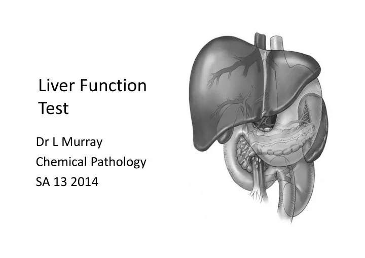
Liver Function Test Dr L Murray Chemical Pathology SA 13 2014 Introduction Anatomy Two lobs, and the lobs consists of lobules Two cell types: Parenchymal cells or hepatocytes (80%) Kupffer cells (Reticuloendothelial system)
Introduction
- Anatomy
– Two lobs, and the lobs consists of lobules – Two cell types:
- Parenchymal cells or hepatocytes (80%)
- Kupffer cells (Reticuloendothelial system)
– Functional unit of the liver is the acinus
- Portal traid (Portal vein, hepatic artery and bile duct) lies at the centre of
the acinus
- Blood flows from portal vein and hepatic artery through sinusoids and
into the hepatic vein, the bile drains in the opposite direction
- Hepatocytes are divided into 3 zones
– Zone 1 (periportal), most blood flow and active in oxidation function, gluconeogenesis and bile formation – Zone 3 (perivascular), least blood flow and active in drug metabolising – Thus disease processes are different in different zones (e.g. most susceptible to viral, toxic and anoxic damage
Structure of the liver
Functions of the liver1
1. Carbohydrate metabolism
- Gluconeogeniesis
- Glycogen synthesis and
breakdown
- Metabolism of lactate
- Galactose metabolism
2. Protein metabolism
- Urea synthesis
3. Lipid metabolism
- Lipoprotein synthesis
- Fatty acid synthesis
- Cholesterol synthesis and
excretion
- Ketogenesis
- Bile acid synthesis
4. Synthetic function
- Synthesis of plasma proteins,
coagulation factors
- Bile acid synthesis
5. Detoxification and excretion
- Bilirubin metabolism
- Foreign compounds (drugs)
6. Metabolism of hormones
- Hydroxylation of vitamin D to
25-OH vitamin D
- Metabolism of peptide
hormones
- Metabolism of steroid
hormones
7. Storage function
- Storage of vit A,D,E,K, B12, iron,
glycogen etc.
Proteins synthesized by liver
1. Plasma proteins except immunoglobulins 2. Proteins involved in heamostasis and fibrinogen
All clotting factors except factor VIII Inhibitors of coagulation – protein C and S, antithrombin III Fibrinolysis factors – plasminogen Antifibrinolysis – alpha2-antiplasmin
3. Hormones
IGF1 Thrombopoietin (regulates platelet production)
4. Apolipoproteins - all except apoB48 5. Prohormones – angiotensinogen 6. Carrier plasma proteins – sex hormone-binding globulin (SHBG), Cortisol-binding proteins (CBG), etc.
Bilirubin metabolism
- About 275 mg of bilirubin is produced daily
- 80% of bilirubin is formed from the breakdown of RBC and its
precursors, the rest is from myoglobin and other heam containing proteins
- In the RES heam is converted to biliverdin and then to
bilirubin, this bilirubin is unconjugated, thus lipid soluble and binds to albumin in the circulation
- Bilirubin binds reversibly to albumin, but in chronic jaundice a
small fraction binds irreversibly and is called gamma-bilirubin
- Bilirubin dissociates from albumin in the liver, this bilirubin is
transported across the hepatocyte membrane by OATP-2
- In hepatocytes, bilirubin is shuttled by ligandin (glutathione-S-
transferase B) and Z protein to rough ER
- In ER bilirubin is conjugated by bilirubin uridine diphosphate
(UDP) glucuronyltransferase (UGT1A1) to form conjugated bilirubin
- UGT1A1 ezyme activity is low at birth and increase after first
10 days of live
- Conjugated bilirubin is actively secreted by multi-drug-
resistant protein 2 (MDRP2) into the bile canaliculi
- Bilirubin entering the intestine:
– Most is oxidised to urobilinogen, which is further oxidised to stercobilin (brown pigment) and urobilin, and excreted in the
- faeces. 20% of urobilinogen is reabsorbed
– Some is deconjugated, then absorbed and re-excreted into the bile (enterohepatic circulation), a small fraction is excreted in the urine because it is water soluble.
- Normally 95% of bilirubin is unconjugated, thus no
bilirubinuria will be present, thus if bilirubinuria is present it is indicative of increased conjugated bilirubin
Bilirubin metabolism
Bile acids
- Bile consists of: bile acids, bile pigments, cholesterol and phospholipids
dissolved in an alkaline electrolyte solution
- Primary bile acids:
– Synthesised from cholesterol – Cholic acids and chenodeoxycholic acid
- Bile salts are stored in the gall bladder and released into the intestine
during digestion
- Most of it is deconjugate by bacterial enzymes and reabsorbed in the
terminal ileum (enterohepatic circulation)
- Secondary bile acids, deoxycholic and lithocholic acids, are formed in the
colon and also reabsorbed again
- Thought that the entire bile acid recycles twice per meal and 6-8 times per
day
- Functions of bile acid:
– Help in the absorption of fat by forming micelles – Activate lipases in intestine – Modulate cholesterol, glucose and triglyceride metabolism by binding to farnesoid X receptor (FRX)
Bile acid metabolism
Conjugation and detoxification or biotransformation and excretion
- Biotransformation:
– Phase I:
- Mediated by microsomal cytochrome P450 oxidase system in ER
- ↑ the polarity of compounds by oxidation
– Phase II:
- The compounds are conjugated making them more water soluble so that
they can be excreted into the urine or bile
- Biotransformation normally lead to detoxification but
sometimes it may lead to toxic or active metabolites
- A lot of cytochrome P450 isoenzymes are present and allelic
variations can lead to intra-individual variations in drug effects
- Other drugs and chemicals may induce (↑ activity) the
enzymes, thus ↑ other compound metabolism
Jaundice
- Yellowish discolouration of
tissue caused by the deposition of bilirubin
- Normally occurs if bilirubin > 35- 40 µmol/L
- Classification
– Pre-hepatic: ↑ production of bilirubin (RBC breakdown) – Hepatic: abnormality or ↓ in conjugation or excretory function of the liver – Post hepatic: Obstruction to the bile flow, intra or extra hepatic
Pre-hepatic Jaundice
- ↑ production of bilirubin due to:
– Destruction of mature RBC (haemolytic anemia) – Ineffective erythopoiesis (pernicious anemia)
- ↑ Unconjugated bilirubin, rarely > 100 µmol/L
(except if associated liver disease)
- Due to ↑ bilirubin production:
– ↑ u-urobilinogen but no u-bilirubin (because unconjugated bilirubin is not water soluble)
Pre-hepatic Jaundice: Causes1
- 1. Ineffective erythropoiesis
– Pernicious anaemia – Thalassaemia
- 2. ↑ RBC breakdown
– Haemolysis
- E.g. Congenital spherocytosis
- Autoimmune haemolysis
– Internal haemorrhage
Hepatic Jaundice
- Due to ↓ capacity of the liver to conjugate and /
- r secrete bile pigments
- Most common cause is hepatitis, destruction of
hepatocytes
- Mixture of conjugate and unconjugated
bilirubinaemia
- Thus urobilinogen and bilirubin may be found in
the urine
Hepatic Jaundice: Causes1
1. Immature conjugating enzymes
Neonatal jaundice
2. Inherited defects in bilirubin metabolism
Gilbert’s syndrome Crigler-Najjar syndrome Rotor syndrome Dubin-Johnson syndrome
3. Hepatic dysfunction
Hepatitis Cirrhosis
4. Drug-induced
Paracetamol Isoniazide
Post-hepatic Jaundice/ Cholestasis
- Cholestasis is when there is interference in
bile flow which may be intra-hepatic or extra- hepatic
- ↑ in conjugated bilirubin
- Bilirubin is detected in the urine, but no
urobilinogen will be present if the obstruction is complete
Post-hepatic Jaundice: Causes1
- 1. Intra-hepatic
Hepatitis Biliary cirrhosis Drugs- anabolic steroids, phenothiazine Hepatic malignancy
- 2. Extra-hepatic
Gallstones Bile duct tumors Compression of bile duct Carcinoma of head of pancreas
Neonatal jaundice2
- Physiological jaundice occurs commonly, due to
immature hepatic conjugating enzymes, ↑ RBC hemolysis and a sterile GIT. Bili is unconjugated and usually < 100 µmol/L
- When to investigate neonatal jaundice
– Present at birth or appears during first 24hr of life – Present beyond 14 days of life – T-bili > 250 µmol/L – Conjugated ↑ bili – Jaundice ass with a clinical picture of disease
Jaundice in the newborn
- Causes of
Unconjugated ↑ bili
– ↑ haemolysis
- Rh incompatibility
- ABO incompatibility
- RBC enzyme defect
– ↓ conjugation
- Crigler-Najjar syndrome
- Hypothyroidism
- Breast milk jaundice
- Causes of Conjugated ↑
bili
– Hemolytic conditions – Hepatitis due to:
- Infection
– Congenital (Rubella, CMV, syphilis) – Acquired (UTI, septicaemia, hepatiis)
- Metabolic disorders
– Alpha1- antitripsin def – Galactosaemia – Tyrosinaemia
- Congenital abnormality
– Biliary atresia
Inherited Hyperbilirubinaemias
- 1. Gilbert’s syndrome
- 2. Crigler-Najjar syndrome
- 3. Dubin-Johnson syndrome
- 4. Rotor’s syndrome
Gilbert’s syndrome
- Benign condition, with an incidence of 5-7% of the
population
- Mild jaundice occur intermittently associated with
illness and starvation
- Other LFT and histology is normal
- Autosomal dominant inheritance, mutation in the
gene leads to ↓ activity of UGT1A1 enzyme (UDP-
glucuronyl transferase enzyme/ Conjugation enzyme)
- Increased unconjugated bilirubin
- It is important to recognise the syndrome to avoid
unnecessary investigations
Crigler-Najjar syndrome
- Rare autosomal recessive condition
– Type 1: Complete absence of UNP-glucuronyl transferase activity, thus complete failure to conjugate bilirubin
- Baby is deeply jaundice in first few days of life
- ↑ unconjugated bilirubin, thus the lipid soluble bilirubin
cross the BBB and deposits in the brain (Kernictus) and death occur in the first year of life
– Type 2: UDP-glucuronyl transferase activity is only reduced
- Jaundice may only appear later on in life
Dubin-Johnson syndrome
- Autosomal recessive disease
- More common in Arabian people
- Mild intermittent jaundice
- Unconjugated and conjugated bilirubin is ↑
- Mutation in the MDRP2, which secretes conjugated
bilirubin from hepatocytes to bile canaliculi
- Other LFT are normal, except bromosulphthalein
(BSP) excretion test
- The liver becomes black due to the accumulation of
brownish pigments
Rotor’s syndrome
- Benign autosomal recessive condition
characterised by fluctuating jaundice due to a failure to secrete bilirubin
- Liver is not pigmented and MDRP2 is normal
- Exact pathogenesis is not fully understood
Assessment of Hepatic Function: Liver function test (LFT)
- The standard LFT are tests to assess liver damage or
dysfunction
- NB: Liver has considerable reserve capacity
- Some of these tests are insensitive to liver damage and may
be normal even when liver damage is present
- LFT are useful to establish
1. If any liver dysfunction is present 2. A specific diagnosis 3. The severity of the liver dysfunction 4. Monitor the progression of disease 5. Assess response to treatment
Liver function tests1
Commonly used tests Function
Transaminases (AST, ALT) Hepatocellular damage Alkaline phosphatase (ALP) Cholestasis Gamma-glutamyl transferase (GGT) Cholestasis, enzyme induction Albumin Synthetic capacity Prothrombin time Synthetic capacity Bilirubin Hepatocellular damage/ cholestasis
Standard liver function tests
- Serum bilirubin
– Not a sensitive test for liver disease – Useful test to monitor the progress of liver disease and to assess response to therapy – Measured as unconjugated (indirect) and conjugated (direct) fractions – Sometimes helpful in diff dx:
- Predominant unconjugated pre-hepatic jaundice
more likely
- Predominantly conjugated cholestasis is likely
Aminotranferases
- Aspartate and alanine aminotransferases (ALT and AST) are
intracellular enzymes
- They are released into the circulation with liver cell damage,
hepatic necrosis and ↑ permeability of hepatocyte membrane
- Sensitive indicators of hepatocellular damage or dysfunction
- Degree of necrosis and inflammation does not correlate with
AST and ALT values
- Very high values are usually due to primary liver disease eg
viral hepatitis
- AST: ALT of > 2 is very suggestive of alcohol-related liver
pathology
Marker of cholestasis: ALP and GGT
- ALP is present in the canalicular plasma membrane
- f hepatocytes and its synthesis is ↑ within hours
after biliary obstruction
- GGT is present in the ER membrane , canalicular
plasma membrane of hepatocytes and biliary epithelial cells
– GGT is non-specific but is a sensitive indicator of cholestasis – Certain drugs and chronic alcohol intake may also ↑ GGT
- ↑ GGT and ALP cholestasis is very likely
Tests of synthetic capacity:
Serum albumin and prothrombin time (PTT)
- ↓ albumin are more indication of chronic liver
disease due to its 20 day half-life and unfortunately albumin concentration can be affected by many
- ther factors
- PTT measure the rate at which prothrombin is
converted to thrombin and is indicative of vit K dependent clotting factors (VII, IX and X) presences, factor VII has the shortest half life (4-6 hrs). Thus prolongation of PTT is an early indicator of impaired liver synthetic capacity, but could also indicate vit K deficiency.
Bile acids
- The sensitivity and specificity of bile acids are
the same as standard LFT, thus its not used routine
- Except in intrahepatic cholestasis of pregnancy
– Where there is intense itching, jaundice and cholestatic features in the third trimester – Bile acids are normal markedly ↑ – ↑ risk of premature delivery and stillbirth
Quantitative LFT1
Less commonly used tests (Quantitative tests) Function Galactose elimination capacity Hepatocyte mass Aminopyrine breath test Microsomal metabolism MEGX formation or Lignocaine clearance test Hepatic blood flow / Microsomal metabolism Bromosulphthlein clearance Hepatic blood flow / Biliary excretion Indocyanine green clearance Hepatic blood flow Caffeine clearance Microsomal metabolism Arterial ketone body ratio
- These tests are mainly used in specialized liver units
Other Liver Function Tests
- High concentrations of Glutathionine-S-
transferase alpha isoenzyme (GST-alpha) is found in the liver.
- Plasma GST-alpha is almost entirely from the
liver
- More sensitive marker of hepatocellular
damage than AST and ALT
- But still only used in research laboratories
Markers of fibrosis
- Fibrosis leads to cirrhosis, thus it would be helpful to
have a marker to diagnose liver fibrosis
- Collagen type I and III deposits in the liver during fibrosis
- Procollagen type III which is cleaved to form the mature
molecule, is useful to monitor patients who are at risk of developing liver fibrosis
- A glycosaminoglycan synthesised in the liver fibrosis,
hyaluronate, has also been used
- Other combination tests are:
– APRI (AST:Platelet ratio) – Fib4 (Platelets, AST and gamma globulin) – Fibrotest (Bilirubin, ALT, GGT, alpha-2 macroglobulin, haptoglobin and apo A1, which are not routinely available)
Tests for diagnosis of specific liver disease
Test Specific liver disease
Alpha feto protein (AFP) Hepatoma, Primary hepatocellular carcinoma
Iron and Iron binding capacity (TIBC) or Transferrin
Haemachromatose Urine Cu and Caeruloplasmin Wilson’s disease Hepatitis A, B, C etc Viral hepatitis Auto-antibodies Autoimmune liver disease Alpha1 antitripsin Alpha1 antitripsin deficiency
Liver Disease
1. Acute hepatitis 2. Acute liver failure 3. Chronic hepatitis Chronic viral hepatitis 4. Autoimmune liver disease Autoimmune hepatitis Primary sclerosing cholangitis Primary biliary cirrhosis 5. Non-Alcoholic Fatty Liver disease (NAFLD)
6.
Liver cirrhosis
Hepatic encephalopathy Ascites Hepatorenal syndrome Endocrine disturbances Portal hypertension Management of cirrhosis
- 7. Alcoholic liver disease
Alcoholic hepatitis
- 8. Drugs and liver
- 9. Malignancy of the liver
biliary tract
- 10. Gallstones
- 1. Acute hepatitis
- Causes
– Infectious : Viral ( Hepatitis A,B,C,D and E) etc – Drugs and toxins
- Typically AST and ALT are > 10 ULN, with a slight increase in ALP and
varying degrees of bilirubineamia
- Hep A:
– 90% of infections may go unrecognized as patients don’t develop jaundice – Symptoms may vary from malaise, anorexia, nausea and fever, dark urine (bilirubin in urine) and the stools may become pale (cholestasis) – Jaundice subsides normally after a few days but some patients will go into a cholestatic phase (↑ GGT and ALP) for weeks
- Recovery
– Hep A and E complete recovery – Hep B, C and D can lead to chronic liver disease
- 10% may progress to chronic liver disease
- <1% may progress to acute liver failure
- The rest may become asymptomatic carriers
2. Acute liver failure
- Characterised by:
– Hepatocellular jaundice (↑ AST and ALT) – Hepatic encephalopathy – ↑ PTT – ↓ Clotting factors II, V, VII, IX and X
- Causes:
– Paracetamol overdose – Viral hepatitis
- Other abnormalities
– Hypoglucaemia (Impaired gluconeogenesis) – Hyponatreamia (Admin of fluids or ↓ free water clearance) – Resp alkalosis (Hyperventilation) / Metabolic acidosis (Hypoxia) – ↓ Ca, Alb and urea (↓ synthesis)
3. Chronic hepatitis
- Def: Clinical or biochemical features of liver
disease (Chronic inflammation) > 6 months
- Chronic viral hepatitis (Hep B, C and D)
– Hep C: 85% will develop chronic infection – Hep B:
- 90% of neonatal hepatitis
- 5-10% of acute infection will become chronic
- 20% of them develop cirrhosis and 5% develop HCC
(Hepatocellular Ca)
– Antiviral therapy may slow progression
4. Autoimmune liver disease
a) Autoimmune hepatitis – Unknown cause, predominantly affects females – Continuing inflammation and necrosis – ↑ AST, ALT and bilirubin as well as ↑ globulins (due to ↑ in gamma globulins [IgG]) – Types:
- Type 1: Anit-smooth muscle antibodies (Abs) (ASMA),
anti-nuclear Abs (ANA), anti-actin Abs (AAA)
- Type 2: Pediatric cases, Anti-liver/kidney microsomal
Abs (LKM)
- Type 3: Cytokeratine Abs
b) Primary sclerosing cholangitis
- Rare chronic conditions, strictures occur in the intra-
and extrahepatic bile duct at any age. It ultimately leads to cirrhosis, liver failure and liver Ca
- LFT: ↑ ALP and GGT
- ↑ Ig G and ANA
- Diagnosis: Characteristic cholangiographic
appearance
- Clinical: Chronic fatigue, jaundice and evidence of
malabsorption (due to strictures of bile duct)
c) Primary biliary cirrhosis
- Chronic, autoimmune liver disease, seen in
middle-age women
- Symptoms: Pruritis, jaundice or non-specific
- LFT: ↑ ALP, GGT and bili (Cholestatic picture)
- ↑ gamma globins due to ↑ IgM
- ↑ Anti-mitochondrial Abs (AMA)
5.
Non-Alcoholic Fatty Liver disease (NAFLD)
- Most common liver disease
- Stages:
– Stage 1: Reversible, benign, fat deposition in hepatocyte – Stage 2: Non-alcoholic steatohepatitis (NASH), inflammation and scaring which can progress to cirrhosis, liver failure and Ca
- Associated with insulin insensitivity, type 2 DM, obesity, drugs
(amiodarone, antivirals, methotrexate, tamoxifen, tetracycline, aspirin etc.)
- In Western society 20-35% of adults have NAFLD, 20-50% of
them have NASH, 20-50% of them will develop cirrhosis
- The fatty change is due to the deposition of ↑ free
fatty acids present in insulin resistance. The further
- xidation of free fatty acids lead to oxidative stress
and ↑ inflammation
- Diagnosis:
– LFT: ↑ AST and ALT, could do tests to establish degree of fibrosis – Ultrasound – Histological examination is diagnostic
- Management:
– ↓ risk factors (weight reduction and exercise, optimal treatment of type 2 DM) – Metformin to ↓ insulin resistance
6. Cirrhosis of the liver1
- Characterized: diffuse hepatic fibrosis and nodular
regeneration
- Causes:
- 1. Alcohol abuse
- 2. Chronic viral hepatitis (Hep B and C)
- 3. NASH
4. Venous obstruction 5. Lupoid hepatitis 6. Cryptogenic hepatitis 7. Autoimmune hepatitis 8. Metabolic disorders 9. Toxins and drugs
- LFT are normal initially due to compensation and
- nly become abnormal in the late stages of disease
- Fibrosis of liver could be monitored by markers like
procollagen III peptide (PIIINP)
- Decompensated cirrhosis:
– Jaundice, ↑ u-urobilinogen and u-bilirubin
– ↓ alb and urea (↓ synthesis)
– ↑ AST, ALT
– ↓ Na (↑ fluid admin and diuretic use) – ↓ K (diuretic use, vomiting and poor diet) – ↓ Mg
Consequences of liver cirrhosis
- 1. Hepatic encephalopathy
- 2. Ascites
- 3. Hepatorenal syndrome
- 4. Portal HT
- 5. Development of hepatic malignancy
- 6. Endocrine dysfunction
Hepatic encephalopathy
- Definition: Impaired mental status and abn
neuromuscular function due to impaired hepatocellular function
- Diagnosis is mainly clinical, EEG changes could be
helpful, ammonia is usually ↑ but is not diagnostic
- Pathogenesis: Multifactorial, toxic products
(ammonia, manganese, natural benzodiazepines, tryptophan) form GIT reaches systemic circulation due to ↓ functioning hepatocytes or portal blood bypassing the liver
- Treatment:
– Removing or treating any precipitating factors (large protein meal, GIT bleeds, infection, ↓ glucose, electrolyte imbalances or drugs) – ↓ protein meals, will ↓ nitrogenous substances absorption – Sterilising GIT by non-absorbable antibiotics (e.g. neomycin) – Adequate energy intake (carbohydrates) – Maintain fluid and electrolyte balance
- Diagram illustrates
the proposed complex feedback mechanisms that can lead to hepatic encephalopathy. BCAA/AAA = branched chain-aromatic amino acids, BZD = benzo diazepines, DA = dopamine, GABA = γ-aminobutyric acid, GLU = glutamic acid, 5HT = serotonin, SCFA = short chain fatty acids.
Ascites1
- Causes:
1. Venous HT
– Cirrhosis of liver – Congestive heart failure – Constrictive pericarditis
2. Hypoalbuminaemia
- Nephrotic syndrome
- Malnutrition (also relevant in cirrhosis)
3. Malignant disease
- Secondary deposits
- Primary mesothelioma
4. Infection
- Tuberculous peritonitis
5. Miscellaneous
- Chylous ascites
- Definition: Ascites is the accumulation of extracellular fluid in
the peritoneal cavity
- Factors involved:
– Retention of Na and water by kidneys – ↓ colloid oncotic pressure (↓ alb) – Portal hypertension – ↑ hepatic lymph production
- Management: Na restriction, diuretics, alb infusion, ascites fluid
withdrawal
- NB: Don’t remove ascites fluid to rapidly, could precipitate renal failure
Fluid analysis Cirrhotic fluid TB or malignancy Protein Concentration < 25 g/L > 25 g/L Serum:ascetic alb ratio > 1.1 < 1.0 Cells per cu mm < 500 > 500 ADA (Adenosine activity) < 30 U/L High (> 30 U/L) in TB
Hepatorenal syndrome
- Definition: Functional ↓ GFR, associated with advanced
liver disease, ascites and encephalopathy
- Kidneys: histologically normal, no ATN (acute tubular
necrosis) present
- Possibility of ↓ GFR, due to vasodilatation of the
splanchnic area (caused by ↑ NO and prostaglandins), leads ↓ circulatory blood volume, activates RAS system, leading to Na retention and vasoconstriction of systemic circulation including renal vessels
- Characterised by: Na retention, u-Na < 10 mmol/l
- Treatment: Albumin infusion and vasopressin analogues
(e.g teripressin)
Endocrine disturbances
- Clinically:
– Men: Impotent, infertile, testicular atrophy, gynecomastia and female distribution of body hair
- Biochemically:
– ↓ Free testosterone, due to ↑ SHBG – ↑ LH with/ without primary testicular failure – ↑ Estrogen due to ↑ peripheral conversion
Portal hypertension
- In cirrhosis the obstruction of the circulation is
caused by the destruction of the liver architecture by fibrosis
- Some features:
– Esophageal varices – GIT bleeds – Haematemesis
Management of liver cirrhosis
- Symptomatic
– Treat underlying cause (alcohol abstinence, metabolic disorders ) – Treat ascites – Adequate nutrition
- Curative treatment is liver transplantation
– MELD (Model for End-stage Liver Disease) score have been used to predict the need for a liver transplantation (Includes bili, creatinine and INR measurements)
7. Alcoholic liver disease
- One of the commonest cause of liver disease worldwide
- Feature which may influence the individuals risk of developing
liver disease – Amount of alcohol consumed – Genetic susceptibility – Gender (F>M) – Nutrition – Associated infections (e.g. Hep B and C)
- Types of alcoholic liver disease:
– Fatty liver (AST, ALT and bili slightly ↑, GGT ↑) – Alcoholic hepatitis – Cirrhosis
Alcoholic hepatitis
- Wide spectrum from asymptomatic to hepatic failure
- LFT: AST/ALT ratio < 2, ALT and AST ↑ (< 10 ULN), GGT
↑
- Liver biopsy is the only way to assess the true degree
- f liver damage
- In asymptomatic patient the markers to indicate
heavy alcohol intake are:
– MCV – GGT – Carbohydrate-deficient (or desialylated) transferrin (Most sensitive)
8. Malignancy of the liver and biliary tract
- Primary
– Hepatocellular Carcinoma
- Associated with Hep B
and C infection, cirrhosis and carcinogens (aflotoxins)
- Alpha fetoprotein (AFP) is
normally ↑
– Cholangio Carcinoma – Angiosarcoma
- Secondary
– Common site for secondaries
9. Drugs causing liver damage1
1. Hepatocellular damage
Paracetamol Alcohol Halothane Methotrexate
2. Cholestasis
Anabolic steroids Naproxen Oral contraceptives Chlordiazepoxide
3. Cirrhosis
Alcohol Methotrexate Methyldopa
4. Chronic cholestasis
Cloxacillin Phenothiazines Tricyclic antidepressants (amitriptyline)
5. Liver tumours
Oral contraceptives Anabolic steriods
6. Chronic hepatitis 7. Hepatic granulomas
Amiodarone Nitrofurantoin Amoxicillin Chlorpromazine Phenylbutazone
- 10. Gallstones
- Common, affect 10-20% of population
- Classified into:
1. Cholesterol: 75%, consists of bili and Ca 2. Pigment: Hard stone (Chr haemolytic disease), Soft stones (Biliary tract infections)
- Predisposing factors
1. Supersaturation of bile with cholesterol – Obesity, ageing, drugs (clofibrate, nicotin) and oestrogen 2. ↓ Bile acid pool 3. Late pregnancy (incomplete emptying of gallbladder) 4. Genetic factors (e.g. Pima Indians)
- Management
– Surgical removal – Chemical (oral treatment with bile acids) or Physical removal
References
1. Swaminathan R (ed). Handbook of Clinical Biochemistry, 2th Ed 2011. World Scientific New Jersey: p 379- 393. 2. Marshall WJ, Bangert SK (eds). Clinical Chemistry, 6th Ed 2008. Mosby Edinburgh: p 93-113; 383-393. 3. Anderson SC, Cockayne S; Clinical Chemistry- Concepts and Applications, Revised ed, p 285-321 4. www.baileybio.com 5. www.britannica.com 6. www.shifa.com 7. www.zuniv.net 8. www.pacbio.com 9. www.kidshealth.org 10. www.arls.gusc.iv 11. www.articlesbeach.com 12. www.radiographics.rsna.org 13. www.medipulse.blogspot 14. You tube videos
1. Liver function tests by Ursula Moore 2. Liver function tests interpretation by Openmichigan