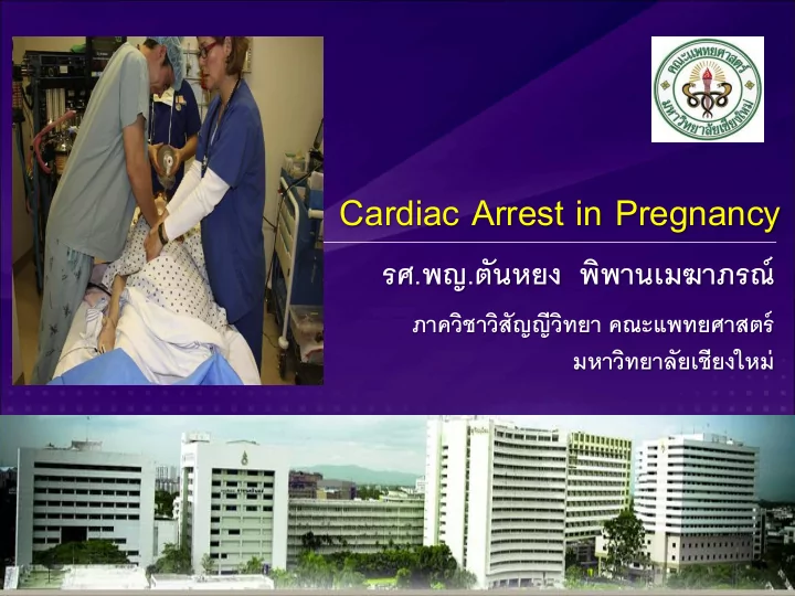

Cardiac Arrest in Pregnancy รศ.พญ.ตันหยง พิพานเมฆาภรณ์ ภาควิชาวิสัญญีวิทยา คณะแพทยศาสตร์ มหาวิทยาลัยเชียงใหม่
Overview • Basic life support (BLS) • Advanced Cardiovascular Life Support (ACLS) • Causes of cardiac arrest • Perimortem cesarean section • Post Cardiac Arrest Care
Maternal cardiac arrest • The overall maternal mortality was calculated at 13.95 deaths per 100,000 maternities. • Incidence: 1:12,000 - 1:20,000 • Poor survival rate (6.9%) • Common causes of cardiac arrest are amniotic fluid embolism, acute myocardial infarction and venous embolism.
Maternal cardiac arrest • Resuscitation of maternal cardiac arrest : - The altered physiologic state induced by pregnancy - The requirement to consider both maternal and fetus issues during resuscitation - Possibility of perimortem cesarean section during resuscitation
Validated obstetric early warning score
BLS Modifications
Basic Life Support Modifications • Patient positioning - important strategy to improve the quality of CPR and resultant compression force and output - effect of aortocaval compression of gravid uterus ( ↓ stroke volume and cardiac output)
Basic Life Support Modifications • Effect of left-lateral tilt - improved maternal hemodynamic parameters (blood pressure, stroke volume, cardiac output) - improved fetal parameters (oxygenation, nonstress test, and heart rate)
Aortocaval compression
Basic Life Support Modifications • No improvement in maternal hemodynamic or fatal parameters with 10 ◦ to 20 ◦ left lateral tilt in patients without cardiac arrest • More aortocaval compression at 15 ◦ left lateral tilt compared to full left lateral tilt • Lateral tilt > 30 ◦ (aortocaval compression)
Basic Life Support Modifications • A tilt ≥ 30 ◦ may not be practical during resuscitation - transmission forces are no longer perpendicular to thorax • Degree of tilt is difficult to estimate (often overestimated)
Manual left uterine displacement (LUD) • Relieve aortocaval compression • Less hypotension and significant reduction in mean ephedrine requirement • Allow high quality chest compression • Easier access for defibrillation and airway management • Left side LUD (preferable)
Recommendations • Continuous manual LUD should be performed on all pregnant women who are in cardiac arrest in which uterus is palpated at or above the umbilicus to relieve aortocaval compression during resuscitation (Class I) • If the uterus is difficult to assess, attempts should be made to perform manual LUD if technically feasible (Class IIb)
Left uterine displacement using 1-handed technique
Left uterine displacement using 2-handed technique
Patient in 30 ◦ left lateral tilt using a firm wedge
Airway • Difficult airway • Lateral tilt position (more difficulty) • Increased risk of aspiration and rapid desaturation • Cricoid pressure (no specific information to support it use) and it should be released if difficult ventilation or poor laryngoscopic view
Breathing • Rapid desaturation - Decreased functional residual capacity - Increased oxygen demand - Increased intrapulmonary shunt • Reduce of ventilation volumes - Elevated diaphragm • Hyperventilation respiratory alkalosis • ( ↑ uterine vasoconstriction and fetal hypoxemia)
Breathing • Prepared to support oxygenation, ventilation, and monitor oxygenation closely
Circulation • Chest compression : similar to nonpregnant (Class IIa) - Position - Rate - Depth
Circulation • The patient should be placed supine for chest compression (Class I) • No literature exam the use of mechanical chest compression and this is not advised at this time
Transporting pregnant women during chest compression • An immediate cesarean delivery may be the best way to optimize the condition of the mother and fetus - This operation should optimally occur at the site of arrest (Class I) - A pregnant patient with in-hospital cardiac arrest should not be transported for cesarean delivery
Defibrillation • The same currently recommended defibrillation protocol should be used in the pregnant patients as in the nonpregnant patients. - No modification of the recommended application of electric shock during the pregnancy (Class I) - The patient should be defibrillated with biphasic shock energy of 120 to 200 J (Class I)
Defibrillation • Compressions should be resumed immediately delivery of the electric shock (Class IIa) • The use of an automated external defibrillator may be considered (Class IIb)
Defibrillation • Anterolateral defibrillator pad placement is recommended as a reasonable default (Class IIa). • The lateral pad/ paddle should be placed under the breast tissue. • The use of adhesive shock electrodes is recommended is to allow consistent electrode placement (Class IIa)
ACLS Modifications
Airway • Changes in airway mucosa (edema, hypersecretion, friability, and hyperemia) • Airway management is more difficult than nonpregnancy • A major cause of maternal morbidity and mortality : failed intubation • The most experienced person should secure and manage
Airway • Bag-mask ventilation with 100% oxygen before intubation is important (Class IIa) • Supraglottic airway devices are acceptable.
Recommendation • Hypoxemia should always be considered as a cause of cardiac arrest. Oxygen reserve are lower and metabolic demand is higher. - Early ventilatory support may be necessary (Class I)
Recommendation • ET tube should be performed by experienced laryngoscopist (Class I) - start with an ET with ID 6.0-7.0 mm (Class I) - optimally no more than 2 laryngoscopy attempt should be made (Class IIa) - supraglottic placement is preferred rescue statergy for failed intubation (Class I)
Recommendation • If attempt airway control fail and mask ventilation is not possible, current guideline for emergency invasive airway access should be followed. • Avoid prolonged intubation attempt to prevent deoxygenation, prolonged interrupted in chest compression, airway trauma, and bleeding) (Class I)
Recommendation • Cricoid pressure is not routinely recommended (Class III) • Continuous waveform capnography - confirming and monitoring correct placement of ET tube - Monitor CPR quality - Adequate chest compression or ROSC (PETCO2> 10 mmHg)
Recommendation • Interruption in chest compression should be minimized during advanced airway placement (Class I)
Circulation • Changes in pharmacokinetics - Increase in glomerular filtration rate - Increase in plasma volume - Decrease in protein binding • However, current recommended drug doses for resuscitation of pregnant patients are similar to adult cardiac arrest (Class IIb)
Circulation • In the setting of cardiac arrest, no medication should be withheld because of concern of teratogenicity (Class IIb) • Current ACLS drugs at recommended doses be used without modifications (Class IIa)
Circulation • The event of difficult peripheral IV access - Intraosseous access in proximal humerus - Ultrasound-assisted peripheral or central venous access • Obtaining IV or intraosseous access above the diaphragm is recommended. - avoid the potentially deleterious effects of vena caval compression
Circulation -Increase the time required for fluids or administered drugs to reach the heart
Defibrillation • Defibrillation should be performed at the recommended ACLS defibrillation doses • Potential harm to the fetus during electrical shock (arcing or electrical burns) • Risk factors for adverse fetal outcomes: current and duration of contact
Defibrillation • The greatest predictor of risk for adverse fetal outcome if the current travels to uterus and amniotic fluid (increased risk of fetal death and burns) • Cardioversion and defibrillation on the external chest are considered safe at all stages of pregnancy.
Defibrillation • If shock is delivered to mother ‘s thorax, there is very low risk of electrical arcing to fetal monitors. • Remove internal or external fetal monitoring is reasonable during maternal cardiac arrest (Class IIb).
Treatment of reversible causes • Obstetric etiologies 1 . Bleeding/ Disseminated Intravascular Coagulation - Expansion of maternal circulating blood volume can mask the sign and symptoms of hemorrhage - Effective quantification of blood loss and awareness of initial changes in maternal vital signs - Crystalloid solutions, blood or blood products or surgical interventions
Treatment of reversible causes 2 . Embolism: coronary, pulmonary, amniotic fluid embolism 2.1 Amniotic fluid embolism -Acute hypotension, cardiovascular collapse and consumptive coagulopathy - Treatment : adequate oxygenation, aggressive restoration of cardiac output, and reverse coagulopathy
Recommend
More recommend