Biochemistry photosynthetic cells within a leaf The ground tissue - - PDF document

biochemistry
SMART_READER_LITE
LIVE PREVIEW

Biochemistry photosynthetic cells within a leaf The ground tissue - - PDF document

10/7/16 Tissue Organization of Leaves : The epidermal barrier in leaves is interrupted by stomata, which allow CO 2 exchange between the surrounding air and the Biochemistry photosynthetic cells within a leaf The ground tissue in a leaf


slide-1
SLIDE 1

10/7/16 1

6) Photosynthesis

Photosynthetic stages and light-absorbing pigments

Biochemistry

  • Prof. Dr. Klaus Heese

Key to labels Dermal Ground Vascular Guard cells Stomatal pore Epidermal cell 50 µm Surface view of a spiderwort (Tradescantia) leaf (LM) (b) Cuticle Sclerenchyma fibers Stoma Upper epidermis Palisade mesophyll Spongy mesophyll Lower epidermis Cuticle Vein Guard cells Xylem Phloem Guard cells Bundle- sheath cell Cutaway drawing of leaf tissues (a) Vein Air spaces Guard cells 100 µm Transverse section of a lilac (Syringa) leaf (LM) (c)

  • Leaf anatomy
  • Tissue Organization of Leaves: The epidermal barrier in leaves is interrupted by

stomata, which allow CO2 exchange between the surrounding air and the photosynthetic cells within a leaf

  • The ground tissue in a leaf is sandwiched between the upper and lower epidermis.

The vascular tissue of each leaf is continuous with the vascular tissue of the stem

(chloroplasts)

This large glucose polymer and the disaccharide fructose are the principle end products of

  • photosynthesis. Both are built of six-carbon sugars. Disaccharide glucose + fructose = sucrose
slide-2
SLIDE 2

10/7/16 2

Photosynthesis: Three of the four stages in photosynthesis occur only during illumination: 1) Absorption of light 2) electron transport leading to formation of O2 from H20, reduction of NADP+ to NADPH, and generation of a proton-motive force pmf 3) Synthesis of ATP, and 4) Conversion of CO2 into carbohydrate, commonly referred to carbon fixation. All four reaction stages of photosynthesis are tightly regulated, coupled and controlled so as to produce the amount of carbohydrate required by the plant. All the reactions in stages 1-3 are catalyzed by proteins in the thylakoid membrane. the enzymes that incorporate CO2 into chemical intermediates and then convert them to starch are soluble constituents of the chloroplast

  • stroma. The enzymes that form sucrose from three-carbon

intermediates are in the cytosol.

Cellular structure of a leaf and chloroplast

Like mitochondria, plant chloroplasts are bounded by a double membrane separated by an intermembrane space. Photosynthesis occurs on the thylakoid membrane, which forms a series of flattened vesicles (thylakoids) that enclose a single interconnected luminal

  • space. The green color of

plants is due to the green color of chlorophyll, all of which is localized to the thylakoid membrane. A granum is a stack of adjacent

  • thylakoids. The stroma is the

space enclosed by the inner membrane and surrounding the thylakoids.

20~50 chloroplasts per cell

slide-3
SLIDE 3

10/7/16 3

Chloroplast stroma Thylakoid space

PSII P680 PSI P700

NADPH ATP

lumen

Stage-1: Absorption of light:

The initial step in photosynthesis is the absorption of light by chlorophylls attached to proteins in the thylakoid membranes. Like the heme component of cytochromes, chlorophylls consist of a porphyrin ring attached to a along hydrocarbon side chain. In contrast to hemes, chlorophylls contain a central Mg2+ ion (rather than Fe atom) and have an additional five-membered ring. The energy of the absorbed light is used to remove electrons form an unwilling donor (water, in green plants), forming oxygen: light 2 H2O ----> O2 + 4 H+ + 4 e- and then to transfer the electrons to a primary electron acceptor, a quinone designated Q, which is similar to CoQ.

Structure of chlorophyll a, the principle pigment that traps light energy.

The CH3 group (green) is replaced by a CHO group in chlorophyll b. In the porphyrin ring (yellow), e- are delocalized among three of the four central rings and the atoms that interconnect

slide-4
SLIDE 4

10/7/16 4

Stage-2: Electron Transport and Generation of a Proton- Motive Force

Electrons move from the quinone primary electron acceptor through a series of electron carriers until they reach the ultimate electron acceptor, usually the

  • xidized form of nicotinamide adenine dinucleotide phosphate (NADP+),

reducing it to NADPH. (NADP is identical in structure with NAD except for the presence of an additional phosphate group. Both molecules gain and lose electrons in the same way. The transport of electrons in the thylakoid membrane is coupled to the movement of protons from the stroma to the thylakoid lumen, forming a pH gradient across the membrane (pHlumen < pHstroma). This process is analogous to generation of a proton-motive force across the inner mitochondrial membrane during electron transport. Thus, the overall reaction of stages 1 and 2 can be summarized as: light 2 H2O + 2 NADP+

  • ---> O2 + 2 H+ + 2 NADPH

Stage-3: Synthesis of ATP

Protons move down their concentration gradient from the thylakoid lumen to the stroma through the F0F1 complex (ATP synthase), which couples proton movement to the synthesis of ATP from ADP and Pi. The mechanism whereby chloroplast F0F1 harnesses the proton-motive force to synthesize the ATP is identical with that used by ATP synthase in the inner mitochondrial membrane and bacterial plasma membrane.

Stage-4: Carbon Fixation

The ATP and NADPH generated by the 2nd and 3rd stages of photosynthesis provide the energy and the electrons to drive the synthesis of polymers of six- carbon sugars from CO2 and H2O. The overall balanced chemical equation is written as: 6 CO2 + 18 ATP4- + 12 NADPH + 12 H2O ---> C6H12O6 + 18 ADP3- + 18 Pi2- + 12 NADP+ + 6 H+ The reactions that generate the ATP and NADPH used in the carbon fixation are directly dependent on light energy; thus stages 1-3 are called the light reactions of photosynthesis. The reactions in stage 4 are indirectly dependent

  • n light energy; they are sometimes called the dark reactions of photosynthesis

because they can occur in the dark, utilizing the suppliers of ATP and NADPH generated by light energy. However, the reactions in stage 4 are not confined to the dark; in fact, they occur primarily during illumination.

Each Photon of Light has a Defined Amount of Energy

Quantum mechanics established that light, a form of electromagnetic radiation, has properties of both waves and particles. When light interacts with matter, it behaves as discrete packets of energy (quanta) called photons. The energy of a photon, e, is proportional to the frequency of the light wave: e = hg, where h is the Planck’s constant (1.58 x 10⁻³⁴ cal s, or 6.63 x 10⁻³⁴ J s) and g is the frequency of the light wave. It is customary in biology to refer to the wavelength of the light wave, l, rather then to its frequency g. with c as the velocity of light (3 x 1010 cm/s in a vacuum) it comes: g = c / l; note hat photons of a shorter wavelength have higher

  • energies. Thus, E = Nhg = Nhc/ l . The energy of light is considerable, as we

can calculate for light with a wavelength of 550 nm (550 x 10-7 cm), typical of sunlight:

E = [(6.02 x 10²³ photons/mol) (1.58 x 10⁻³⁴ cal s) (3 x 1010 cm/s) ] / (550 x 107cm) = 51,881 cal/mol

  • r about 52 kcal/mol. This is enough energy to synthesize several moles of ATP

from ADP and Pi, if all the energy were used for this purpose.

slide-5
SLIDE 5

10/7/16 5

Photosystems comprise a Reaction Center and Associated Light-Harvesting Complexes

The absorption of light energy and its conversion into chemical energy occurs in multiprotein complexes called photosystems. Found in all photosynthetic

  • rganisms, both eukaryotic and prokaryotic, photosystems consist of two closely

linked components: a reaction center, where the primary events of photosynthesis

  • ccur, and an antenna complex consisting of numerous protein complexes, termed

light-harvesting complexes (LHCs), which capture light energy and transmit it to the reaction center. Both reaction centers and antennas contain tightly bound light- absorbing pigment molecules. Chlorophyll a is the principal pigment involved in photosynthesis, being present in both reaction centers and antennas. In addition to chlorophyll a, antennas contain other light-absorbing pigments: chlorophyll b in vascular plants and carotenoids in both plants and photosynthetic bacteria. Carotenoids consist of long hydrocarbon chains with alternating single and double bonds; they are similar in structure to the visual pigment retinal, which absorbs light in the eye. The presence of various antenna pigments, which absorb light at different wavelengths, greatly extends the range of light that can be absorbed and used for photosynthesis.

Photosystems comprise a Reaction Center and Associated Light-Harvesting Complexes

One of the strongest pieces of evidence for the involvement of chlorophylls and carotenoids in photosynthesis is that the absorption spectrum of these pigments is similar to the action spectrum of photosynthesis. The latter is a measure of the relative ability of light of different wavelengths to support photosynthesis. When chlorophyll a (or any other molecule) absorbs visible light, the absorbed light energy raises the chlorophyll a to a higher energy (excited) state. This differs from the ground (unexcited) state largely in the distribution of electrons around the C and N atoms of the porphyrin ring. Excited states are unstable and return to the ground state by one of several competing processes. For chlorophyll a molecules dissolved in organic solvents such as ethanol, the principal reactions of light (fluorescence and phosphorescence) and thermal emission (heat). When the same chlorophyll a is bound to the unique protein environment of the reaction center, dissipation of excited-state energy occurs by a quite different process that is the key to photosynthesis.

The rate of photosynthesis is greatest at wavelengths of light absorbed by three pigments

The action spectrum of photosynthesis in plants, that is, the ability of light of different wavelengths to support photosynthesis, is shown in black. Absorption spectra for three photosynthetic pigments present in the antennas of plant photosystems are shown in color. Each absorption spectrum shows how well light of different wavelengths is absorbed by one of the pigments. A comparison of the action spectrum with the individual absorption spectra suggests that photosynthesis at 680 nm is primarily due to light absorbed by chlorophyll a; at 650 nm to light absorbed by chlorophyll b, and at shorter wavelengths to light absorbed by chlorophyll a and b and by carotenoid pigments, including b-carotene

Photoelectron transport, the primary event in photosynthesis, from energized reaction-center chlorophyll a produces a charge separation After absorption of a photon of light, one of the excited special pair of chlorophyll a molecules in the reaction center (left) donates an electron to a loosely bound acceptor molecule, the quinone Q, on the stromal surface of the thylakoid membrane, creating an essentially irreversible charge separation across the membrane (right). The electron cannot easily return through the reaction center to neutralize the positively charged chlorophyll a.

slide-6
SLIDE 6

10/7/16 6

Photoelectron transport, the primary event in photosynthesis, from energized reaction-center chlorophyll a produces a charge separation The absorption of a photon of light of wavelength = 680 nm by chlorophyll a increases by its energy by 42 kcal/mol (the first excited state). Such an energized chlorophyll a molecule in a plant reaction center rapidly donates an electron to an intermediate acceptor, and the electron is rapidly passed on to the primary electron acceptor, quinone Q, on the stromal surface of the thylakoid membrane. This light-driven electron transfer, called photoelectron transport, depends on the unique environment of both the chlorophylls and the acceptor within the reaction center. Photoelectron transport, which

  • ccurs nearly every time a photon is absorbed, leaves a positive charge on the

chlorophyll a close to the luminal surface and generates a reduced, negatively charged acceptor (Q-) near the stromal surface. The Q- produced by photoelectron transport is a powerful reducing agent with a strong tendency to transfer an electron to another molecule, ultimately to NADP+. The positively charged chlorophyll a+, a strong oxidizing agent, attracts an electron from an electron donor on the luminal surface to regenerate the original donor on the luminal surface to regenerate the original chlorophyll a. In plants, the oxidizing power of four chlorophyll a+ molecules is used, by way of intermediates, to remove four electrons form 2 H2O molecules bound to a site on the luminal surface to form O2. 2 H2O + 4 chlorophyll a+

  • --> 4 H+ + O2 + 4 chlorophyll a

These potent biological reductants and oxidants provide all the energy needed to drive all subsequent reactions of photosynthesis: electron transport, ATP synthesis, and CO2 fixation. Photoelectron transport, the primary event in photosynthesis, from energized reaction-center chlorophyll a produces a charge separation Chlorophyll a also absorbs light at discrete wavelengths shorter than 680 nm. Such absorption raises the molecule in several higher excited states, which decay within 10-12 seconds (1 picosecond, ps) to the first excited state with loss of the extra energy as heat. Because photoelectron transport and the resulting charge separation occur only from the first excited state of the reaction-center chlorophyll a, the quantum yield – the amount of photosynthesis per absorbed photon – is the same for all wavelengths of visible light shorter (and, therefore, of higher energy) than 680 nm.

Light-Harvesting Complexes increases the Efficiency of Photosynthesis

Energy transfer from light-harvesting complexes to associated reaction center in photosystem I of cyanobacteria

The multiprotein light-harvesting complex binds 90 chlorophyll molecules (white and blue) and 31 other small molecules, all held in a specific geometric arrangement for optimal light absorption. Of the six chlorophyll molecules (green) in the reaction center, two constitute the special-pair chlorophylls (ovals) that can initiate photoelectron transport when excited (blue arrows). Resonance transfer of energy (red arrows) rapidly funnels energy from absorbed light to one of two “bridging” chlorophylls (blues) and thence to chlorophylls in the reaction center.

Energy is transferred from the LHC chlorophyll molecules to reaction-center chlorophylls by resonance energy transfer

Taking home message:

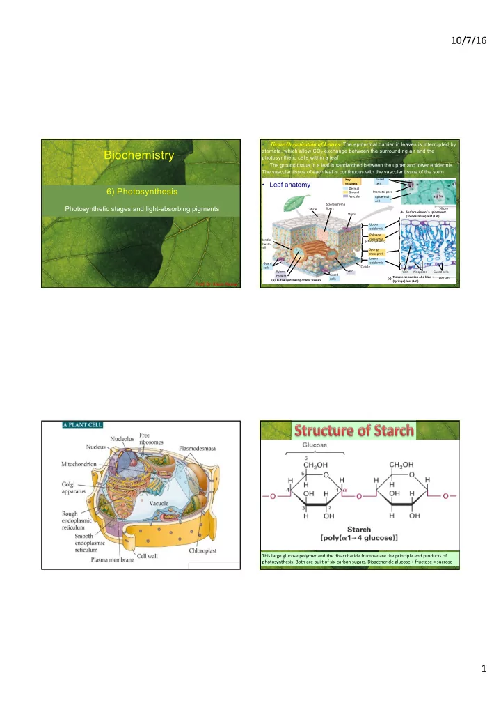
  • The principle end products of photosynthesis in plants are oxygen and polymers of

six-carbon sugars (starch and sucrose)

  • The light capturing and ATP-generating reactions of photosynthesis occur in the

thylakoid membrane located within chloroplasts. The permeable outer membrane and inner membrane surrounding chloroplasts do not participate in photosynthesis.

  • In stage 1 of photosynthesis, light is absorbed by chlorophyll a molecules bound to

reaction-center proteins in the thylakoid membrane. The energized chlorophylls donate an electron to a quinone on the opposite side of the membrane, creating a charge separation. In green plants, the positively charged chlorophylls then remove electrons from water, forming oxygen.

  • In stage 2, electrons are transported from the reduced quinone via carriers in the

thylakoid membrane until they reach the ultimate electron acceptor, usually NADP+, reducing it to NADPH. Electron transport is coupled to movement of protons across the membrane from the stroma to the thylakoid lumen, forming a pH gradient (proton- motive force pmf) across the thylakoid membrane.

  • In stage 3, movement of protons down their electron-chemical gradient through F0F1

complexes powers the synthesis of ATP from ADP and Pi.

slide-7
SLIDE 7

10/7/16 7

Taking home message:

  • In stage 4, the ATP and NADPH generated in stage 2 and 3 provide energy and the

electrons to drive the fixation of CO2 and synthesis of carbohydrates. These reactions

  • ccur in the thylakoid stroma and cytosol.
  • Associated with each reaction center are multiple light-harvesting complexes (LHCs),

which contain chlorophylls a and b, carotenoids, and other pigments that absorb light at multiple wavelengths. Energy is transferred from the LHC chlorophyll molecules to reaction-center chlorophylls by resonance energy transfer.

3-D structure of photosynthetic reaction center form the purple bacterium Rhodobacter spheroides

Top: the L subunit (yellow) and M subunit (white) each form five transmembrane a helices and have a very similar structure overall; the H subunit(light blue) is anchored to the membrane by a single transmembrane a helix. A fourth subunit (not shown) is a peripheral protien that binds to the exoplasmic segments of the other subunits. Bottom: within each reaction center is a special pair of bacteriochlorophyll a molecules (green), capable of initiating photoelectron transport; two voyeur chlorophylls (purple); two pheophytins (dark blue), and two quinones, QA and QB (orange). QB is the primary electron acceptor during photosynthesis.

Linear electron flow in plants, which requires both chloroplast photosystem, PSI and PSII

Blue arrows indicate flow of electrons; red arrows indicate proton movement. LHCs are not shown. Left: in the PSII reaction center, two sequential light-induced excitations, of the same P680 chlorophylls result in reduction of the primary electron acceptor QB to QH2. on the luminal side of PSII, electrons removed from H2O in the thylakoid lumen are transferred to P680+, restoring the reaction-center chlorophylls to the ground state and generating O2. Center: the cytochrome bf complex then accepts electrons from QH2, coupled to the release of two protons into the lumen. Operation of a Q cycle in the cytochrome bf complex translocates additional protons across the membrane to the thylakoid lumen, increasing the proton-motive force pmf generated. Right: in the PSI reaction center, each electron released from light-excited P700 chlorophylls moves via a series of carriers in the reaction center to the stromal surface, where soluble ferredoxin (an Fe-S protein) transfers the electron to FAD and finally to NADP+, forming NADPH. P700+ is restored to its ground state by addition of an electron carried from PSII via the cytochrome bf complex and plastocyanin, a soluble electron carrier. The pmf generated by linear electron flow from PSII to NADP-FAD reductase powers ATP synthesis by the F0F1 complex.

In linear system, ATP synthesis and NADPH generation Only PSII generates a H+ gradient, No H+ gradient in PSI

Cyclic electron flow in the single photo-system of purple bacteria

Blue arrows indicate flow of electrons; red arrows indicate proton movement. Left: energy funneled by an associated LHC (not illustrated here) energizes one of the special-pair chlorophylls in the reaction center. Photoelectron transport from the energized chlorophyll, via pheophytin (Ph) and quinone A (QA), to quinone B (QB) forms the semiquinone Q-. and leaves a positive charge on the chlorophyll. Following absorption of a second photon and transfer of a second electron to the semiquinone, it rapidly picks up two protons from the cytosol to form QH2. Center: After diffusing through the membrane and binding to the Qo site on the exoplasmic face

  • f the cytochrome bc1 complex, QH2 donates two electrons and simultaneously gives up two protons to the external medium,

generating a proton-motive force pmf (H+exoplasmic > H+cytosolic). Electrons are transported back to the reaction-center chlorophyll via a soluble cytochrome, which diffuses in the periplasmic space. Operation of a Q cycle in the cytochrome bc1 complex pumps additional protons across the membrane to the external medium, as in mitochondria. The pmf is used by the F0F1 complex to synthesize ATP and , as in other bacteria, to transport molecules in and out of the cell.

In cycling system, only ATP synthesis

slide-8
SLIDE 8

10/7/16 8

Electron flow and O2 evolution in chloroplast PSII

The PSII reaction center, comprising two integral proteins, D1 and D2, special-pair chlorophylls (P680), and other electron carriers, is associated with an oxygen-evolving complex on the luminal surface. Bound to the three extrinsic proteins (33, 23, and 17 kDa) of the oxygen-evolving complex are four manganese ions (red), a Ca2+ ion (blue), and a Cl- ion (yellow). These bound ions function in the splitting of H2O and maintain the environment essential for high rates of O2 evolution. Tyrosine-161 (Y161) of the D1 polypeptide conducts electrons from the Mn ions to the oxidized reaction-center chlorophyll (P680+), reducing it to the ground state P680.

A single PSII absorbs a photon and transfers an electron four times to generate one O2 = 4 photons generate 1 O2

Dark-adapted chloroplasts were exposed to a series of closely spaced, sort (5 µs) pulses of light that activated virtually all the PSIIs in the preparation. The peaks in O2 evolution occurred after every fourth pulse, indicating that absorption of four photons by one PSII is required to generate each O2 molecule. Because the dark-adapted chloroplasts were initially in a partially reduced state, the peaks in the O2 evolution occurred after flashes 3, 7, and 11.

Distribution of multiprotein complexes in the thylakoid membrane and the regulation of linear versus cyclic electron flow

Top: in sunlight, PSI and PSII are equally activated, and the photosystems are organized in state I. in this arrangement, light- harvesting complex II (LHCII) is not phosphorylated and is tightly associated with the PSII reaction center in the grana. As a result, PSII and PSI can function in parallel in linear electron flow. Bottom: when light excitation of the two photosystems is unbalanced, LHCII becomes phosphorylated, dissociates from PSII, and diffuses into the unstacked membranes, where it associates with the PSI and its permanently associated LHCI. In this alternative supramolecular organization (state II), most of the absorbed light energy is transferred to PSI, supporting cyclic electron flow and ATP production but no formation of NADPH and thus no CO2 fixation. PC = plastocyanin.

No O2 generation No NAPDH ---> No Carbon fixation

Taking home message:

  • In the single photosystem (of purple bacteria), cyclic electron flow from light-excited

chlorophyll a molecules in the reaction center generates a proton-motive force pmf, which is used mainly to power ATP synthesis by the F0F1 complex in the plasma membrane.

  • Plants contain two photosystems PSI and PSII, which have different functions and are

physically separated in the thylakoid membrane. PSII splits H2O into O2. PSI reduces NADP+ to NADPH. Cyanobacteria have two analogous photosystems.

  • In chloroplasts, light energy absorbed by light-harvesting complexes (LHCs) is

transferred to chlorophyll a molecules in the reaction centers (P680 in PSII and P700 in PSI).

  • Electrons flow through PSII via the same carriers that are present in the bacterial
  • photosystems. In contrast to the bacterial system, photochemically oxidized P680+ in

PSII is regenerated to P680 by electrons derived from the splitting of H2O with evolution of O2.

  • In linear electron flow, photochemically oxidized P700+ in PSI is reduced, regenerating

P700, by electrons transferred from PSII via the cytochrome bf complex and soluble

  • plastocyanin. Electrons lost from P700 following excitation of PSI are transported via

several carriers ultimately to NADP+, generating NAPDH.

slide-9
SLIDE 9

10/7/16 9

Taking home message:

  • In contrast to linear electron flow, which requires both PSII and PSI, cyclic electron

flow in plants involves only PSI. In this pathway, neither NADPH nor O2 is formed, although a proton-motive force pmf is generated.

  • The pmf generated by photoelectrons transport in plant and bacterial photosystems is

augmented by operation of the Q cycle in cytochrome bf complexes associated with each of the photosystems.

  • Reversible phosphorylation and dephosphorylation of the PSII light-harvesting

complex control the functional organization of the photosynthetic apparatus in thylakoid membranes. State I favors linear electron flow, whereas state II favors cyclic electron flow.

CO2 Metabolism during Photosynthesis

The initial reaction that fixes CO2 into organic compounds

In this reaction, catalyzed by ribulose 1,5-bisphosphate carboxylase (rubisco), CO2 condenses with the five-carbon sugar ribulose 1,5 bisphosphate. The products are two molecules of 3-phosphoglycerate. enzyme: rubisco (via stoma)

The pathway of carbon during photosynthesis

Top: six molecules of CO2 are converted into two molecules of glyceraldehyde 3 phosphate. These reactions, which constitute the Calvin cycle,

  • ccur in the stroma of the chloroplast. Via phosphate/triosephosphate antiporter, some glyceraldehyde 3-phosphate is transported to the cytosol

in exchange for phosphate. Bottom: in the cytosol, an exergonic series of reactions converts glyceraldehyde 3-phosphate to fructose 1,6- bisphosphate and, ultimately, to the disaccharide sucrose. Some glyceraldehyde 3-phosphate (not shown here) is also converted to amino acids and fats, compounds essential to plant growth. The fixation of 6 CO

2 molecules and the net formation of 2 glyceraldehyde 3-phosphate molecules

require the consumption of 18 ATPs and 12 NADPHs, generated by the light-requiring process of phosphosynthesis.

slide-10
SLIDE 10

10/7/16 10

  • fructose and glucose
  • tastes sweet

– fruit, vegetables, grains

  • table sugar is refined

sugarcane and sugar beets

  • brown, white, powdered

Sucrose

CO2 fixation and photorespiration

(In C3 plants,) these competing pathways are both initiated by ribulose 1,5-bisphosphate carboxylase (rubisco), and both utilize ribulose 1,5-bisphosphate . CO2 fixation, pathway 1, is favored by high CO2 and low O2 pressures; photorespiration, pathway 2, occurs at low CO2 and high O2 pressures (that is under normal atmospheric conditions). Phosphoglycolate is recycled via a complex set of reactions that take place in peroxisomes and mitochondria, as well as chloroplasts. The net result: for every two molecules of phosphoglycolate formed by photorespiration (four C atoms), one molecule of 3-phosphoglycerate is ultimately formed and recycled, and one molecule of CO2 is lost.

In C3 plants, photorespiration recycles the C in PO4-glycolate produced in the

  • xygenation reaction of Rubisco to glycerate with the release of 1/4 of the C as
  • CO2. The reactions involve 3 different organelles.

This is called photo- respiration because it only occurs in the light (when Rubisco is active), releases CO2 (in the mito= chondria) and uses O2 (in the peroxi- somes)

Leaf anatomy of C4 plants and the C4 pathway. a) In C4 plants, bundle sheath cells line the vascular bundles containing the xylem and phloem. Mesophyll cells, which are adjacent to the substomal air spaces, can assimilate CO2 into four-carbon molecules at low ambient CO2 and deliver it to the interior bundle sheath cells. Bundle sheath cells contain abundant chloroplasts and are the sites of phosphosynthesis and sucrose synthesis. Sucrose is carried to the rest of the plant via the phloem. In C3 plants, which lack bundle sheath cells, the Calvin cycle operates in the mesophyll cells to fix CO2. b) the key enzyme in the C4 pathway is phosphoenolpyruvate carboxylase, which assimilate CO2 to form oxaloacetate in mesophyll cells. Decarboxylation of malate or other C4 intermediates in bundle sheath cells releases CO2, which enters the standard Calvin cycle.

HCO3-

C4 plants: Spatial separation of CO2 fixation and Calvin Cycle (Rubisco reaction)

slide-11
SLIDE 11

10/7/16 11

slide-12
SLIDE 12

10/7/16 12

(PEP) vacuole vacuole guard cells guard cells a) b)

a) Water and salts enter the xylem through the roots. Water is lost by evaporation, mainly through the leaves, creating a suction pressure that draws the water and dissolved salts upward through the xylem. The phloem is used to conduct dissolved sucrose, produced in the leaves, to

  • ther parts of the plant. b) enlarged view illustrates the mechanism of sucrose flow in a higher plant. Sucrose is actively transported from

mesophyll cells into companion cells, and then moves through plasmodesmata into the sieve-tube cells that constitute the phloem vessels. The resulting increase in osmotic pressure within the phloem causes water carried in xylem vessels to enter the phloem by osmotic flow. Root cells and other nonphotosynthetic cells remove sucrose from the phloem by active transport and metabolize it. This lowers the osmotic pressure in the phloem, causing water to exit the phloem. These differences in osmotic pressure in the phloem between the source and the sink of sucrose provide the force that drives sucrose through the phloem.

Schematic diagrams of the two vascular systems – xylem and phloem – in higher plants, showing the transport of water (blue) and sucrose (red)

slide-13
SLIDE 13

10/7/16 13

Biomass-Energy Algae Biomass

Taking home message:

  • In the Calvin cycle, CO2 is fixed into organic molecules in a series of reactions that
  • ccur in the chloroplast stroma. The initial reaction, catalyzed by rubisco, forms a 3-

carbon intermediate. Some of the glyceraldehyde 3-phosphate generated in the cycle is transported to the cytosol and converted to sucrose.

  • The light-dependent activation of several Calvin cycle enzymes and other

mechanisms increase fixation of CO2. in the light.

  • In C3 plants, much of the CO2 fixed by the Calvin cycle is lost as the result of

photorespiration, a wasteful reaction catalyzed by rubisco that is favored at low CO2 and high O2 pressures.

  • In C4 plants, CO2 is fixed initially in the outer mesophyll cells by reaction with
  • phosphoenolpyruvate. The 4-carbon molecules, so generated, are shuttled to the

interior bundle sheath cells, where the CO2 is released and then used in the Calvin

  • cycle. The rate of photorespiration in C4 plants is much lower than in C3 plants.
  • Sucrose from photosynthetic cells is transported through the phloem to

nonphotosynthetic parts of the plant. Osmotic pressure differences provide the force that drives sucrose transport.

slide-14
SLIDE 14

10/7/16 14

  • The light reactions and chemiosmosis: the organization of

the thylakoid membrane

LIGHT REACTOR

NADP+ ADP ATP NADPH

CALVIN CYCLE

[CH2O] (sugar)

STROMA (Low H+ concentration) Photosystem II LIGHT

H2O CO2

Cytochrome complex

O2

H2O O2 1

1⁄2

2 Photosystem I Light THYLAKOID SPACE (High H+ concentration) STROMA (Low H+ concentration) Thylakoid membrane ATP synthase Pq Pc Fd NADP+ reductase NADPH + H+ NADP+ + 2H+ To Calvin cycle ADP P ATP 3 H+ 2 H+ +2 H+ 2 H+

In linear system, ATP synthesis and NADPH generation Only PSII generates a H+ gradient, No H+ gradient in PSI lumen

The Importance of Photosynthesis: A Review

  • A review of photosynthesis

Light reactions:

  • Are carried out by molecules in the

thylakoid membranes

  • Convert light energy to the chemical

energy of ATP and NADPH

  • Split H2O and release O2 to the

atmosphere Calvin cycle reactions:

  • Take place in the stroma
  • Use ATP and NADPH to convert

CO2 to the sugar G3P

  • Return ADP, inorganic phosphate, and

NADP+ to the light reactions

O2 CO2 H2O Light Light reaction Calvin cycle

NADP+ ADP ATP NADPH + P 1 RuBP 3-Phosphoglycerate

Amino acids Fatty acids Starch (storage) Sucrose (export)

G3P Photosystem II Electron transport chain Photosystem I Chloroplast

END