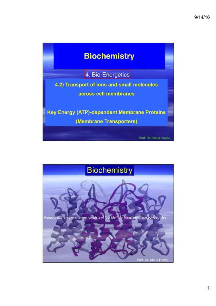
biochemistry
play

Biochemistry 4. Bio-Energetics 4.2) Transport of ions and small - PDF document

9/14/16 Biochemistry 4. Bio-Energetics 4.2) Transport of ions and small molecules across cell membranes Key Energy (ATP)-dependent Membrane Proteins (Membrane Transporters) Prof. Dr. Klaus Heese Biochemistry Aquaporin, the water channel,


  1. 9/14/16 Biochemistry 4. Bio-Energetics 4.2) Transport of ions and small molecules across cell membranes Key Energy (ATP)-dependent Membrane Proteins (Membrane Transporters) Prof. Dr. Klaus Heese Biochemistry Aquaporin, the water channel, consists of four identical transmembrane polypeptides Prof. Dr. Klaus Heese 1

  2. 9/14/16 Relative permeability of a pure phospholipid bilayer to various molecules A bilayer is permeable to small hydrophobic molecules and small uncharged polar molecules, slightly permeable to water and urea, and essentially impermeable to ions and to large polar molecules. Membrane Transport Proteins Gradients are indicated by triangles with the tip pointing toward lower concentration, electrical potential, or both. 1: pumps utilize the energy released by ATP hydrolysis to power movement of specific ions (red circles) or small molecules against their electrochemical gradient. 2: Channels permit movement of specific ions (or water) down their electrochemical gradient; they can also be controlled by e.g. ligand binding or phosphorylations etc : Transporters, which fall into three groups, facilitate movement of specific small molecules or ions. Uniporters transport a single type of molecule down its concentration gradient (3A). Cotransport proteins (symporters (3B) and antiporters (3C) catalyze the movement of one molecule against its concentration gradient (black circle), driven by movement of one or more ions down an electrochemical gradient (red circles). Differences in the mechanisms of transport by these three major classes of proteins account for their varying rates of solute movement. Transporters can also depend on ATP. 2

  3. 9/14/16 Cellular uptake of glucose mediated by GLUT proteins exhibit simple enzyme kinetics and greatly exceeds the calculated rate of glucose entry solely by passive diffusion The initial transport rate for the substrate S into the cell catalyzed by e.g. GLUT1: v=Vmax/(1+Km/[S]) The initial rate of glucose uptake (measured as micromoles per milliliter of cells per hour) in the first few seconds is plotted against increasing glucose concentration in the extracellular medium. In this experiment, the initial concentration of glucose in the cells is always zero. Both, GLUT1, expressed by erythrocytes, and GLUT2, expressed by liver cells, greatly increase the rate of glucose uptake (red and orange curves) at all external concentrations. Like enzyme-catalyzed reactions, GLUT-facilitated uptake of glucose exhibits a maximum rate (Vmax). The Km is the concentration at which the rate of glucose uptake is half maximal. GLUT2, with a Km of about 20 mM, has a much lower affinity for glucose than GLUT1, with a Km of about 1.5 mM. 3

  4. 9/14/16 Model of Uniport Transport by GLUT1 In one conformation, the glucose-binding site faces outward; in the other, the binding site faces inward. Binding of glucose to the outward-facing site (step-1) triggers a conformational change in the transporter that results in the binding site facing inward toward the cytosol (step-2). Glucose then is released to the inside of the cell (step 3). Finally, the transporter undergoes the reverse conformational change, regenerating the outward-facing binding site (step 4). If the concentration of glucose is higher inside the cell than outside, the cycle will work in reverse (step-4 ---> step-1), resulting in net movement of glucose form inside to out. The actual confomrational changes are probably smaller than those depicted here. Operational model for the 2-Na + /1-glucose symporter Simultaneous binding of Na + and glucose to the conformation with outward-facing binding sites (step-1) generates a second conformation with inward-facing site (step-2). Dissociation of the bound Na + and glucose into the cytosol (step-3) allows the protein to revert to its original outward-facing conformation (step-4), ready to transport additional substrate. 4

  5. 9/14/16 Liposomes containing a single type of transport protein are very useful in studying functional properties of transport proteins Here, all the integral proteins of the erythrocyte membrane are solubilized by a nonionic detergent, such as octylglucoside. The glucose uniporter GLUT1 can be purified by chromatography on a column containing a specific antibody and then incorporated into liposomes made of pure phospholipids. ATP-Powered Pumps and the Intracellular Ionic Environment 5

  6. 9/14/16 The 4 classes of ATP-powered transport proteins - (1) P-class pumps are composed of a catalytic alpha subunit which becomes phosphorylated as part of the transport cycle. A beta subunit, present in some of these pumps, may regulate (regulatory subunit) transport. Operational model of the Ca 2+ ATPase in the SR membrane of skeletal muscle cells Only one of the two catalytic alpha subunits of this P-class pump is depicted. E1 and E2 are alternative conformations of the protein in which the Ca 2+ -binding sites are accessible to the cytosolic and exoplasmic faces, respectively. An ordered sequence of steps (1-6) is essential for coupling ATP hydrolysis and the transport of Ca 2+ ions across the membrane. In this figure ~P indicates a high-energy acyl phosphate bond; -P indicates a low-energy phosphoester bond. Because the affinity of Ca 2+ for the exoplasmic-facing sites in E2, this pump transports Ca 2+ uni- directionally from the cytosol to the SR lumen. 6

  7. 9/14/16 Structure of the catalytic alpha subunit of the muscle Ca 2+ ATPase (a) 3-D models of the protein in the E1 state based on the structure determined by X-ray crystallography. There are 10 transmembrane alpha helices, 4 of which (green) contain residues that site-specific mutagenesis studies have identifies as participating in Ca 2+ binding. The cytosolic segment forms 3 domains: the nucleotide binding domain (orange) the phosphorylation domain (yellow) and the actuator domain (pink) that connects 2 of the membrane-spanning helices. (b) Hypothetical model of the pump in E2 state, based on a lower resolution structure determined by electron microscopy of frozen crystals of the pure protein. Note the differences between the E1 and E2 states in the confirmations of the nucleotide- binding and actuator domains; these changes probably power the conformational changes of the membrane-spanning alpha helices (green) that constitute the Ca 2+ -binding site, converting them form one in which the Ca 2+ -binding sites are accessible to the cytosolic face (E1 state) to one in which they are accessible to the exoplasmic face (E2 state). Operational model of the Na + /K + ATPase in the plasma membrane Only one of the two catalytic alpha subunits of this P-class pump is depicted. It is not known whether only one or both subunits in a single ATPase molecule transport ions. Ion pumping by the Na + /K + ATPase (very imporant in neurons) involves phosphorylation, dephosphorylation and conformational changes similar to those in the muscle Ca 2+ ATPase, in this case, hydrolysis of the E2-P intermediate powers the E2--->E1 conformational change and concomitant transport of two ions (K + ) inward. Na + ions are indicated by red circles; K + ions by purple squares; high energy acyl phosphate bond by ~P; low-energy phosphoester bon by -P. 7

  8. 9/14/16 The 4 classes of ATP-powered transport proteins - (2) V-class pumps do not form phosphoprotein intermediates and transport only protons. V/F-class structures are similar and contain similar proteins, but none of their subunits are related to the P-class pumps. V-class pumps couple ATP hydrolysis to transport of protons against a concentration gradient. Effect of proton pumping by V-class ion pumps on H + concentration gradients and electric potential gradients across cellular membranes. (a) If an intracellular organelle contains only V-class pumps, proton pumping generates an electric potential across the membrane, luminal-side positive, but no significant change in the intraluminal pH. (b) if the organelle also contains Cl - channels, anions passively follow the pumped protons, resulting in an accumulation of H + ions (low luminal pH) but no electric potential across the membrane. 8

  9. 9/14/16 The 4 classes of ATP-powered transport proteins - (3) change of ? pH [ion] e - -potential electromotive force (emf) F-class pumps do not form phosphoprotein intermediates and transport only protons. V/F-class structures are similar and contain similar proteins, but none of their subunits are related to the P-class pumps. F-class pumps operate in the reverse directions (compared to V-class) to utilize energy in a proton concentration or electrochemical gradient to synthesize ATP. The 4 classes of ATP-powered transport proteins - (4) All members of the large ABC superfamily of proteins contain 2 transmembrane (T) domains and 2 cytosolic ATP-binding (A) domains, which couple ATP hydrolysis to solute movement. These core domains are present as separate subunits in some ABC proteins, but are eventually fused to a single polypeptide in other ABC proteins. 9

Download Presentation
Download Policy: The content available on the website is offered to you 'AS IS' for your personal information and use only. It cannot be commercialized, licensed, or distributed on other websites without prior consent from the author. To download a presentation, simply click this link. If you encounter any difficulties during the download process, it's possible that the publisher has removed the file from their server.

Recommend


More recommend