background
play

Background ECMO is a life support machine replaces the function of - PDF document

Lesson 19 University of Bergamo Engineering and Management for Health FOR CHRONIC DISEASES MEDICAL SUPPORT SYSTEMS LESSON 19 Extra Corporeal Membrane Oxygenation (ECMO) Ettore Lanzarone May 13, 2020 Background ECMO is a life support


  1. Lesson 19 University of Bergamo Engineering and Management for Health FOR CHRONIC DISEASES MEDICAL SUPPORT SYSTEMS LESSON 19 Extra Corporeal Membrane Oxygenation (ECMO) Ettore Lanzarone May 13, 2020 Background 
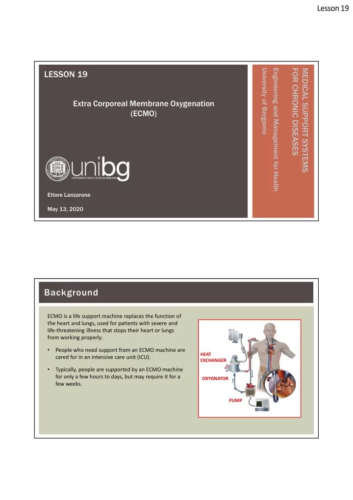
ECMO is a life support machine replaces the function of the heart and lungs, used for patients with severe and life-threatening illness that stops their heart or lungs from working properly. • People who need support from an ECMO machine are HEAT cared for in an intensive care unit (ICU). EXCHANGER • Typically, people are supported by an ECMO machine for only a few hours to days, but may require it for a OXYGNATOR few weeks. PUMP

  2. Lesson 19 Background ECMO is used to help people whose: • Lungs cannot provide enough oxygen to the body even when given extra oxygen • Lungs cannot get rid of carbon dioxide even with help from a mechanical ventilator HEAT • Heart cannot pump enough blood to the body EXCHANGER ECMO may also be used to support people with heart or OXYGNATOR lung disease that cannot be cured while they wait for an organ transplant ( e.g., new heart and/or lungs ). PUMP Background The ECMO machine is connected to a patient through plastic tubes ( cannula ). These tubes are placed in large veins and arteries in the legs, neck or chest. The ECMO machine pumps blood from the patient’s body to an artificial lung (oxygenator) that adds oxygen HEAT to it and removes carbon dioxide. EXCHANGER Patients on ECMO are also given a medication to thin the OXYGNATOR blood (e.g., heparin) so it does not clot. Any patient connected to an ECMO machine in the ICU is PUMP also connected to monitors. They measure heart rate, blood pressure, oxygen and carbon dioxide levels, and level of heparin.

  3. Lesson 19 Background Extra corporeal circuits started to appear about in 1950, due to the technical difficulties of the oxygenation function. Example of experiment in dog in 1940 Background The core of the ECMO machine is the oxygenator. We will focus on dimensioning and controlling the oxygenator. The dimensioning is an off-line procedure (the patient receives a given oxygenator with fixed characteristics), while the control on-line (perfusion conditions can be varied during ECMO). Thus, we will address: • Dimensioning • Control

  4. Lesson 19 Oxygenator The idea is to mimic the lung. They have a modular structure, whose exchange unit is the alveolus, a sort of bag containing the air coming from the bronchi, around which a network of capillaries is wrapped. Mass exchange (oxygenation and CO 2 extraction of from blood are regulated by the Fick's law). Its rigorous formulation of this law is differential terms, but dimensioning and control of oxygenators its expression it in finite terms is sufficient:  p  partial N k R Oxygenator  p  partial p p N k 1 2 R N with: flow ml   N Gas flow N transferred from the system 1 to the system 2  area min m 2 is directly proportional to the partial pressure difference of the same between the two compartments and is  p partial  mmHg inversely proportional to a flow resistance, which is represented by the obstacle between the two chambers. Systems 1 and 2 may contain gases, liquids or a liquid and a gas, but the law remains unchanged.

  5. Lesson 19 Oxygenator It is possible to identify an analogy between Fick's law and Ohm's law, with the following correspondences:  N (gas flow) I (electrical current)  p (partial pressure difference)  V (potential difference)  R (resistance to flow) R (electrical resistance) Oxygenator The blood comes into contact with the alveolar atmosphere through a partition wall, consisting of several biological membranes. In an average adult, the lungs have an exchange surface at the alveolar level of about 50-70 m 2 . Likely, in oxygenators, the exchange surface can be very resized. 1. The lungs are sized to guarantee the exchange even during the performance of a high physical activity, while oxygenators must guarantee the oxygenation of a patient in the ICU, whose oxygen demand is limited; already for this, the surface can be reduced about 5 times. Notice that, without making intense efforts, even people with only one lung can survive. 2. The composition of the gas that oxygenates the blood is a parameter that can be directly set and controlled, therefore it is possible to use a mixture whose oxygen content is much higher than that of the alveolar atmosphere. An increase of  p produces an increase in the flow and consequently an increase in the quantity of oxygen exchanged.

  6. Lesson 19 Oxygenator The composition of the external air is: • 20% of O 2 (also depending on the altitude) • 80% of N 2 • 0.04% of CO 2 During the inspiration phase, the air coming from the external environment is mixed with the “exhaust gases” of metabolic processes, with a different composition. Thus, the average composition of the alveolar air is characterized by intermediate conditions: • 13% of O 2 • 6% of CO 2 The amount of N 2 remains unchanged as it is not exchanged. These values in the alveolar air correspond to: • pO 2 = 100 mmHg • pCO 2 = 40 mmHg The relative average partial pressures in the venous blood reaching the lungs are: • pO 2 = 40 mmHg • pCO 2 = 46 mmHg Oxygenator Therefore, in the case of oxygen, the  pO 2 between gas and blood is about 60 mmHg. This corresponds to the required 50-70m 2 . To reduce the area, a supply of air with higher pO 2 can be exploited (up to the pure oxygen with pO 2 = 760mmHg, i.e., the atmospheric pressure). Thus,  pO 2 can be increased up to  pO 2 = 760 mmHg – 40 mmHg = 720 mmHg. This value is about 10 times greater than the physiological one, so it requires a surface 10 times smaller. An area of about 7 m 2 will suffice, which is the size used in several oxygenators.

  7. Lesson 19 Oxygenator Oxygen and carbon dioxide are transported by blood in two ways: • Dissolved in plasma • Linked to hemoglobin (Hb) in red cells Only the first way would not be enough to guarantee the oxygenation to tissues and this explain the role of Hb in red cells. On average, the amount of Hb in the blood at the physiological Ht = 45% is about 14g per 100 ml and each gram of Hb is able to bind, at its maximum capacity, 1.36 ml of oxygen. In this situation it is said that arterial blood is 100% saturated. Actually, arterial blood is 98-99% saturated. Oxygenator Abacus that relates pO 2 , Sat and concentration of linked O 2 ( which is about equal to the total amount ). These relationships depend on Ht and temperature.

  8. Lesson 19 Oxygenator Oxygenator Considering these numbers, the exchanged quantities are: With a blood flow of 5 l/min the overall quantities are: Unbalancing is compensated by eliminating H 2 0 in the urine. The dissolved quantity is negligible and thus neglected.

  9. Lesson 19 Oxygenator Now, let us focus on the resistance to flow R . Gases meet a series of resistances as they pass. As for O 2 : • the first barrier is the alveolar membrane, a semipermeable membrane that opposes its passage offering a certain resistance; • the oxygen molecule passes from the alveolar membrane to the capillary membrane through the interstitial fluid; • before reaching the blood, it also crosses the membrane that constitutes the capillary wall. C0 2 does the opposite route. Oxygenator We start in a disadvantaged situation compared to the lung, at least from the geometric point of view. • Technology is not yet able to produce membranes as thin and permeable as nature does. • It is impossible to build conducts for blood passage with transversal sections of radius of 8-10  m (like the capillaries). By schematizing an oxygenator as a container in which a membrane separates the gas compartment from the blood, we can rewrite Fick's law as ( resistances in series ):  t m = thickness of membrane p  partial t s = thickness of the blood film N k t t D m = diffusion coefficient of the gas through the membrane  m s D s = coefficient of diffusion of the gas in the blood D D m s Note that the CO 2 diffusion coefficient in blood is 6 to 15 times greater than the O 2 one.

  10. Lesson 19 Oxygenator Classification of oxygenators There exist three types of oxygenators, which are listed in historical order of appearance: 1. bubble oxygenators; 2. film oxygenators; 3. membrane oxygenators: i. real membrane oxygenators (continuous membrane); ii. hybrid oxygenators (microporous membrane). Nowadays, the most used oxygenator is the membrane one and in particular the hybrid one, even if in terms of market percentage the bubbling oxygenators still cover a good slice. They are used because they damage the blood less, even if they are more expensive. They are used also in the ECMO ( “M” is for Membrane ). Oxygenator  p The design goal is to reduce the flow resistance R:  partial N k • t t Thin membranes to reduce t m  m s • Reduce the thickness t b of blood film D D m s Two technological solutions allow to keep the thickness t b low: • tube bundle : a set of ducts (capillaries) in which one of the two phases flows, separated from the other phase that laps the outer surface of the capillaries • parallel flat plates : blood flows in the space delimited by two plates, while gas flows in the contiguous interspace between two other plates. With these configurations, the thickness of the blood film is defined as the semi-distance between two membranes.

Download Presentation
Download Policy: The content available on the website is offered to you 'AS IS' for your personal information and use only. It cannot be commercialized, licensed, or distributed on other websites without prior consent from the author. To download a presentation, simply click this link. If you encounter any difficulties during the download process, it's possible that the publisher has removed the file from their server.

Recommend


More recommend