
SLIDE 1
3/26/2019 1
Slide 1 JSOMTC, SWMG(A)
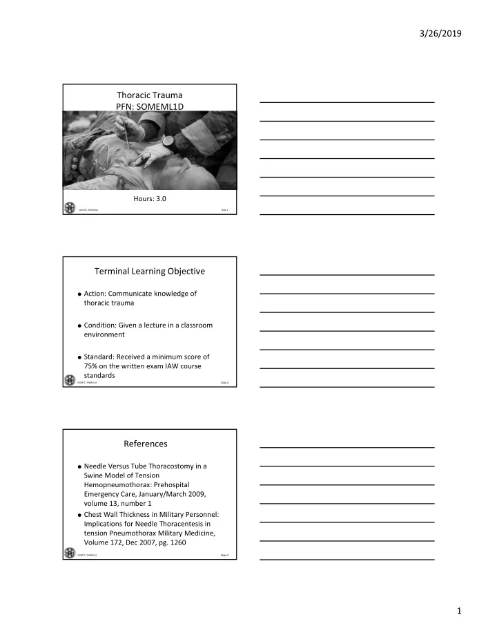
Thoracic Trauma PFN: SOMEML1D
Hours: 3.0
Slide 2
JSOMTC, SWMG(A)
Terminal Learning Objective
Action: Communicate knowledge of
thoracic trauma
Condition: Given a lecture in a classroom
environment
Standard: Received a minimum score of
75% on the written exam IAW course standards
Slide 3
JSOMTC, SWMG(A)
References
Needle Versus Tube Thoracostomy in a
Swine Model of Tension Hemopneumothorax: Prehospital Emergency Care, January/March 2009, volume 13, number 1
Chest Wall Thickness in Military Personnel: