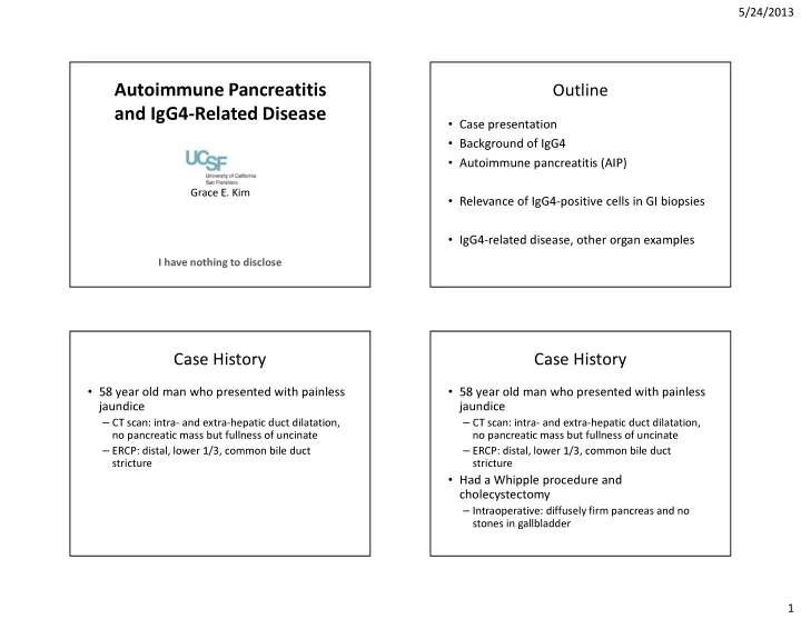

5/24/2013 Autoimmune Pancreatitis Outline and IgG4-Related Disease • Case presentation • Background of IgG4 • Autoimmune pancreatitis (AIP) Grace E. Kim • Relevance of IgG4-positive cells in GI biopsies • IgG4-related disease, other organ examples I have nothing to disclose Case History Case History • 58 year old man who presented with painless • 58 year old man who presented with painless jaundice jaundice – CT scan: intra- and extra-hepatic duct dilatation, – CT scan: intra- and extra-hepatic duct dilatation, no pancreatic mass but fullness of uncinate no pancreatic mass but fullness of uncinate – ERCP: distal, lower 1/3, common bile duct – ERCP: distal, lower 1/3, common bile duct stricture stricture • Had a Whipple procedure and • Had a Whipple procedure and cholecystectomy cholecystectomy – Intraoperative: diffusely firm pancreas and no – Intraoperative: diffusely firm pancreas and no stones in gallbladder stones in gallbladder 1
5/24/2013 Cellular stroma Storiform fibrosis No discrete mass lesion Obliterative phlebitis Perineural inflammation 2
5/24/2013 Periductal inflammation Lymphocytes, plasma cells, eosinophils Numerous IgG4-positive plasma cells >50 IgG4+ plasma cells/HPF 3
5/24/2013 Common bile duct inflammation IgG4+/IgG+ plasma cells = >40% Transmural inflammation of gallbladder Lymphoplasmacytic inflammation 4
5/24/2013 >50 IgG4+ plasma cells/HPF IgG4+/IgG+ plasma cells = >40% Chronic pancreatitis My diagnosis IgG4-related disease Autoimmune pancreatitis type 1 (IgG4-related pancreatitis ) IgG4-related cholecystitis 5
5/24/2013 Chronic pancreatitis Immunoglobulin G (IgG) • Most abundant immunoglobulin (75-80%) • Four subclasses – IgG4 accounts for 3-6% of total serum IgG Figure from http://course1.winona.edu/kbates/Immunology/images/figure_09_37.jpg Serum IgG4 concentration Started with…and where we are now • Upper limit of normal is variable – 86 mg/dL at UCSF – 121 mg/dL in another lab • Elevated serum IgG4 – >135 mg/dL • Sensitivity of 97%; specificity of 79.6% in diagnosing IgG4- related disease • Patients with allergic disorders, receiving allergen immunotherapy, parasitic disease, pemphigus, variety of pulmonary disorders, and reported in rheumatoid arthritis 6
5/24/2013 IgG4-related disease (IgG4-RD) IgG4-RD major organ manifestations • Diffuse or mass forming fibro-inflammatory Pancreas (prototype) Biliary tree Gallbladder condition rich in IgG4-positive plasma cells Liver Orbit/periorbital Sinus/nose – Diagnosis based on combination of Salivary gland Lymph nodes Thyroid • Clinical, imaging, serology, histopathology and Mediastinum Aorta Pericardium immunohistochemistry Lung Retroperitoneum Kidney • Multiorgan disease can be synchronous or Pituitary Meninges Peripheral nerve evolve metachronously over months to years Skin Breast Prostate *Stomach *Bowel *Mesentery *Spleen * Suspected, not confirmed N Eng J Med. 2012;366:539-51. Two main features of IgG4-RD How to count IgG4-positive plasma cells • At x40 objective lens (HPF) 1. Characteristic histologic appearance – Use printed photographs of the same microscopic a. Dense lymphoplasmacytic infiltrate field b. Fibrosis, least focally in a storiform pattern – Direct counting under microscope, but – NOT by “eyeballing” c. Obliterative phlebitis • Find “hot spots” (most intense IgG4+ foci) 2. Elevated number of IgG4-positive plasma • Count three HPF then calculate average cells in tissue • Use same fields on IgG stain to calculate IgG4+/IgG+ ratio 7
5/24/2013 Non-IgG4-RD cases with increased IgG4+ cells Pitfalls • Inflammatory conditions • Diagnosing IgG4-RD because of excessive (Abundant plasma cells, so high numbers of IgG4+ plasma cells) – Primary sclerosing cholangitis (23%) emphasis on elevated serum IgG4 level – Inflammatory bowel disease – 10% of pancreatic adenocarcinoma – Autoimmune atrophic gastritis, oral inflammatory diseases, anti-neutrophilic cytoplasmic antibody-associated – 20% of cholangiocarcinoma vasculitits, rheumatoid arthritis, Rosai-Dorfman disease, Hashimoto’s thyroiditis, Castleman’s disease, pulmonary • Overreliance on IgG4+ plasma cells in tissue abscess, splenic sclerosing angiomatoid nodular transformation, perforating collagenosis, inflammatory myofibroblastic tumor, and Rhinosinusitis • Malignancy – Pancreatobiliary cancer – Lymphoma N Engl J Med 2012;366:539-551. Threshold IgG4-to-IgG ratio does not equate to IgG4-RD • Powerful tool – >40% comprehensive cutoff value in any organ • Sensitivity 94.4%, specificity of 85.7% – Useful particularly when abundance of plasma cells For histology Ratio is also alone, use a must the term. . . Figure from Mod Pathol. In J Rhematol 2012; 2012:580814. 2012;25:1181-1192. 8
5/24/2013 Balance histologic criteria and Autoimmune pancreatitis (AIP) diagnosis of IgG4-related disease • Stringent criteria provides high specificity AIP type 1 AIP type 2 Infiltrate Dense predominantly Dense predominantly lymphoplasmacytic – In appropriate clinical context lymphoplasmacytic infiltrate with neutrophilic infiltration Pancreatic Without epithelial damage and With destruction of duct epithelium • Elevated serum IgG4 ducts patent lumen by neutrophilic granulocytes • Other organ manifestation of disease ( granulocytic epithelial lesion ) Lobules Involving and replacing acinar Patchy involvement commonly admixed • Diagnosis can be made with lower IgG4+ count and tissue with neutrophils Fibrosis Storiform fibrosis , most Less prominent, limited to pancreas IgG4+/IgG+ ratio prominent in peripancreatic fat • But clinical features must be correlated with Vein Obliterative phlebitis Obliterative phlebitis rarely seen histopathologic criteria IgG4 stain Abundant positive plasma cells Scant to no positive plasma cells Modified Honolulu consensus Why? Management Reason to subtype • AIP type 1 is responsive to corticosteroid • Recurrence risk – Remission in 3 months (87-98%) – AIP type 1 • High 3-year relapse rate (6-59%) • AIP type 2 • Predictor of relapse – Has been observed to improve with – Presence of IgG4-related cholangitis/proximal duct corticosteroids involvement – Spontaneous resolution • Whipple procedure – Decrease risk of relapse (2.7-28%) – Does not eliminate risk of relapse – AIP type 2 does not relapse J Gastrointest Surg;2013;17:8990906. 9
5/24/2013 Clinical profile of autoimmune pancreatitis Classical imaging findings of pancreas • Computed tomography (CT) scan AIP type 1 AIP type 2 Mean age ~62 years old ~48 years old – Diffuse enlargement and effacement of the usual Male 61-91% 44-74% lobular appearance Elevated serum • Endoscopic retrograde IgG4 level 41-76% 0-17% cholangiopancreatography (>135 mg/dL) – Diffuse or long segments of irregular narrowing Other organ Biliary, salivary, involvement retroperitoneal, of the main pancreatic duct kidney Prevalence of Absent; 2-6% Present; 16-30% IBD Tumefactive mass Serum IgG4 in pancreatic disease Clinical presentation and radiographic Cut off >140 mg/dL: Sensitivity (76%), Specificity (93%), PPV (36%) appearance mimics pancreatic carcinoma and Cut off >280 mg/dL: Sensitivity (53%), Specificity (53%), PPV (75%) leads to pancreatic resection. In a surgical series of resections for “chronic pancreatitis” AIP Normal Pancreatic Benign Acute Chronic Miscell- pancreas cancer pancreatic pancreatitis pancreatitis aneous • tumor AIP represented about 20% of Whipple resections Elevated serum IgG4 in 7% of non-AIP patients • Only 33% had a discrete mass on CT scan 9.6% of patients with pancreatic cancer (13/135, 9.6%) Figure taken from Am J Gastroenterol 2007;102:1646-1653. 10
5/24/2013 Dense ductocentric inflammation with no Autoimmune pancreatitis type 1 epithelial duct damage and patent lumen Histologic features Infiltrate Dense predominantly lymphoplasmacytic infiltrate Pancreatic Without epithelial damage and ducts patent lumen Lobules Involving and replacing acinar tissue Fibrosis Storiform fibrosis , most prominent in peripancreatic fat Vein Obliterative phlebitis IgG4 stain Abundant positive plasma cells Inflammatory infiltrate Inflammatory infiltrate • Lymphocytes Can also include – Diffuse T-cells; CD3-, mild to moderate CD4-, CD8-positive eosinophils, scattered – Germinal center B-cells macrophages, and • Plasma cells rare neutrophils 11
5/24/2013 Variable lobular involvement Storiform fibrosis Venulitis Obliterative phlebitis 12
5/24/2013 Lymphoid aggregate? EVG stain facilitates Pitfalls of obliterative phlebitis Pitfalls of obliterative phlebitis • Can be observed in chronic pancreatitis and pancreatic adenocarcinoma • Lymphoid aggregate adjacent to artery mistaken for obliterative phlebitis 13
Recommend
More recommend