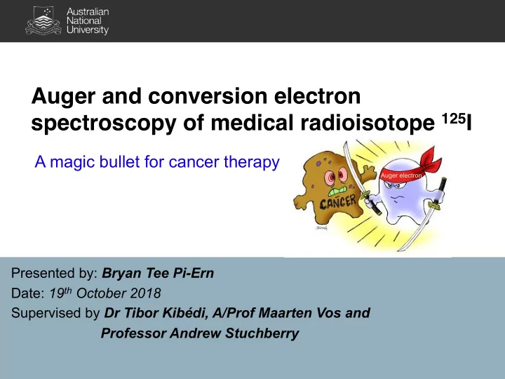

Auger and conversion electron spectroscopy of medical radioisotope 125 I A magic bullet for cancer therapy Auger electron Presented by: Bryan Tee Pi-Ern Date: 19 th October 2018 Supervised by Dr Tibor Kibédi, A/Prof Maarten Vos and Professor Andrew Stuchberry
Strand of human DNA α β 2
Strand of human DNA Auger e - α β 3
Decay scheme of 125 I 4
Decay scheme of 125 I Conversion coefficient ! = # $ # % e - (Internal conversion) or γ (Gamma decay) 5
Decay scheme of 125 I Mixing ratio of the M1+E2 transition is known to be small ( δ << 1). e - (Internal conversion) or γ (Gamma decay) 6
Decay scheme of 125 I Penetration effects !(M1) = ! 0 ((1)(1 + * 1 + + * 2 + 2 ) Where λ = penetration parameter e - (Internal conversion) or γ (Gamma decay) 7
X-ray and Auger transitions X-ray transition Auger transition K Auger yield ω K + a K = 1 E K ≈ E K - - E E L 3 L 3 M 2 M 2 K fluorescence yield E K ≈ E K - E L 3 L 3 E M 2 E E L 3 L 3 E K E K *Atomic notations: K = 1s 1/2 , L 3 = 2p 3/2 , M 2 = 3p 1/2
Vacancy cascade X v Resulting in heaps of Auger electrons v Energy range: a few eV to 30 keV (for 125 I case) X K Note: X = X-ray transition, A = Auger transition 9
Computational model - BriccEmis v Calculate the Auger and X-ray spectra using a Monte Carlo approach v Transition probabilities from Evaluated Atomic Data Library (EADL) (Perkins 1991) v Transition energies are calculated using the relativistic self-consistent-field Dirac Fock method, using RAINE code (Band 2002) L L 10 Probability [/100 decays] 8 6 4 KLL KLL M M,N,O 2 KLM N KLX 0 20 22 24 26 28 30 32 34 36 Energy [keV] 10
Project Aims v Measure an accurate Auger yield from medical radioisotope 125 I Approach Determine the nuclear parameters ( λ and δ) I. II. Measure the Auger to conversion electrons intensity ratios. III. Deduce the absolute intensity of Auger electrons from the conversion coefficients. 11
Project Aims v Measure an accurate Auger yield from medical radioisotope 125 I v Test and benchmark the model 12
Source preparation v Monolayer of 125 I on top of a gold substrate 125 I Au(111) 125 I Au(111) 13
High-energy electrostatic spectrometer HV hemisphere (Positive high voltage) Vos et al. (2000) Hemispherical electron 2D detector energy analyser (Close to ground potential) 14
High-energy electrostatic spectrometer HV hemisphere (Positive high voltage) Energy range: 2 keV to 40 keV Vos et al. (2000) Hemispherical electron 2D detector energy analyser (Close to ground potential) 15
Conversion electron measurements 16000 L 1 Brabec et al. 1982 14000 Miura et al. 1986 Casey et al. 1968 12000 2018 Present work. 2017 10000 Counts 8000 Resolution ≈ 5 eV 6000 4000 L 2 2000 L 3 0 30400 30600 30800 31000 31200 Energy [eV] 16
Conversion electron line shapes Voigt profiles Main peak 17
Nuclear parameters determination 125 I 35.4925 keV M1+E2 λ =5.0(21) ANU data only δ =0.0000(84) v Chi-square fitting method 2018ANU L2/L1 v Reduced χ 2 = 0.63 2018ANU L3/L1 v λ = 5.0(21), δ = 0.0000(84) 2018ANU M1/L1 Experiment Atomic shell Present work Literature 6.68(14) [12] 100/(1+ T ot ) 6.55(13) [13] 2018ANU M2/M1 12.95(28) [15] a T ot 14.25(64) [8] 0.80(5) [16] K/ (1 + T ot ) 0.804(10) [17] L/ (1 + T ot ) 0.11(2) [16] M/ (1 + T ot ) 0.020(4) [16] 2018ANU M3/M1 11.78(18) a [15] K 11.90(31) [8] L 1.4(1) [18] K/L 12.3(25) [10] L/M 5.21(26) [9] 2018ANU M2/L1 M/N 4.87(20) [9] 1:0.089(4):0.024(2) [7] 1:0.106(22):0.041(2) [10] L 1 : L 2 : L 3 1:0.085(2):0.019(2) 1:0.082(4):0.019(3) [8] 1:0.095(2):0.023(5) [9] L 1 : M 1 1: 0.204(7) - 2018ANU N1/M1 1:0.092(5):0.044(3) [8] M 1 : M 2 : M 3 1:0.094(6):0.022(7) 1:0.101(5):0.030(5) [9] L 1 : M 2 1:0.0173(26) - − 0.5 0 0.5 1 1.5 2 M 1 : N 1 1:0.179(20) 1:0.214(6) [9] α Exp / α Fit a Corrected ω K to 0.875 18
Nuclear parameters determination 125 I 35.4925 keV M1+E2 λ =0.2(7) All data δ =0.0132(71) 1952Bo16 K/(1+Tot) 1952Bo16 L/(1+Tot) v Chi-square fitting method 1952Bo16 M/(1+Tot) 1959Na06 K/L 1959Na06 K/M 1959Na06 K/N v Reduced χ 2 = 1.55 1965Ge04 L1/L2 1965Ge04 L1/L3 1969Ka08 Tot 1969Ka08 K v λ = 0.2(7), δ = 0.0132(71) 1969Ca01 L1/L2 1969Ca01 L1/L3 1969Ca01 K/L 1970Ma51 K/(1+Tot) Experiment 1979CoZG Tot Atomic shell 1979CoZG K Present work Literature 1979CoZG L2/L1 6.68(14) [12] 1979CoZG L3/L1 100/(1+ T ot ) 6.55(13) [13] 1979CoZG M2/M1 12.95(28) [15] a 1979CoZG M3/M1 T ot 14.25(64) [8] 1982Br16 L/M 0.80(5) [16] 1982Br16 M/N K/ (1 + T ot ) 0.804(10) [17] 1982Br16 L1/L2 1982Br16 L1/L3 L/ (1 + T ot ) 0.11(2) [16] 1982Br16 M1/M2 M/ (1 + T ot ) 0.020(4) [16] 11.78(18) a [15] 1982Br16 M1/M3 K 1982Br16 M2/M3 11.90(31) [8] 1982Br16 M1/N1 L 1.4(1) [18] 1990Iw04 γ − ray K/L 12.3(25) [10] 1992ScZZ γ − ray L/M 5.21(26) [9] 1999Sa55 L M/N 4.87(20) [9] 2018ANU L2/L1 1:0.089(4):0.024(2) [7] 2018ANU L3/L1 1:0.106(22):0.041(2) [10] L 1 : L 2 : L 3 1:0.085(2):0.019(2) 2018ANU M1/L1 1:0.082(4):0.019(3) [8] 2018ANU M2/M1 1:0.095(2):0.023(5) [9] 2018ANU M3/M1 L 1 : M 1 1: 0.204(7) - 2018ANU M2/L1 1:0.092(5):0.044(3) [8] M 1 : M 2 : M 3 1:0.094(6):0.022(7) 2018ANU N1/M1 1:0.101(5):0.030(5) [9] L 1 : M 2 1:0.0173(26) - − 0.5 0 0.5 1 1.5 2 M 1 : N 1 1:0.179(20) 1:0.214(6) [9] α Exp / α Fit a Corrected ω K to 0.875 19
KLL Auger electron measurements 10000 10000 (b) L 1 Conversion (a) KLL Auger 3500 9000 9000 Experiment KL 2 L 3 BrIccEmis 8000 8000 KL 1 L 3 + KL 2 L 2 3000 7000 7000 6000 6000 Counts KL 1 L 2 KL 3 L 3 KL 1 L 1 2500 5000 5000 4000 4000 2000 3000 3000 2000 2000 1500 1000 1000 21600 22000 22400 22800 30450 30600 Energy (eV) Energy [eV] 20
KLL Auger electron measurements 10000 10000 (b) L 1 Conversion (a) KLL Auger 3500 Auger to conversion ratio 9000 9000 Experiment KL 2 L 3 BrIccEmis underestimated by 20% 8000 8000 KL 1 L 3 + KL 2 L 2 3000 7000 7000 6000 6000 Counts KL 1 L 2 KL 3 L 3 KL 1 L 1 2500 5000 5000 4000 4000 2000 3000 3000 2000 2000 1500 1000 1000 21600 22000 22400 22800 30450 30600 Energy (eV) Energy [eV] 21
KLM Auger electron measurements 5000 (b) L 1 Conversion (a) KLM Auger 12000 12000 Experiment 4900 KL 3 M 1 KL 3 M 2 + KL 2 M 4 KL 2 M 5 + KL 3 M 3 BrIccEmis 11000 11000 4800 10000 10000 KL 1 M 2 9000 9000 KL 1 M 1 KL 1 M 3 4700 Counts KL 2 M 1 KL 1 M 4,5 KL 2 M 2,3 8000 8000 4600 7000 7000 4500 6000 6000 5000 5000 4400 4000 4000 25600 26000 26400 26800 30450 30600 Energy (eV) Energy (eV) 22
KLM Auger electron measurements 5000 (b) L 1 Conversion (a) KLM Auger 12000 12000 Auger to conversion ratio Experiment 4900 KL 3 M 1 KL 3 M 2 + KL 2 M 4 KL 2 M 5 + KL 3 M 3 BrIccEmis underestimated by 20% 11000 11000 4800 10000 10000 KL 1 M 2 9000 9000 KL 1 M 1 KL 1 M 3 4700 Counts KL 2 M 1 KL 1 M 4,5 KL 2 M 2,3 8000 8000 4600 7000 7000 4500 6000 6000 5000 5000 4400 4000 4000 25600 26000 26400 26800 30450 30600 Energy (eV) Energy (eV) 23
K fluorescence yield determination Karttunen (1969) ω K + a K = 1 Tolea (1974) Singh (1990) 0.13 0.87 30% 5% Ozdemir (2002) 0.15 Yashoda (2005) 0.85 10% 2% Present (2017) 0.12 0.88 20% EADL (1991) 3% 0.65 0.7 0.75 0.8 0.85 0.9 ω K ω K 24 24
K fluorescence yield determination Karttunen (1969) ω K + a K = 1 Semi-empirical Tolea (1974) value (Schonfeld, 1996) Singh (1990) 0.13 0.87 30% 5% Ozdemir (2002) 0.15 Yashoda (2005) 0.85 10% 2% Present (2017) 0.12 0.88 20% EADL (1991) 3% 0.65 0.7 0.75 0.8 0.85 0.9 ω K ω K 25 25
K fluorescence yield global fit curve Semi-empirical fit typical error: 3% Data compilation up to 2017 (ANU Summer research, 2017) 26
What to do next? v Quantify the Auger electrons with energy < 1 keV v Effects of electron shake-off following internal conversion and Auger transition (the tails) v Atomic structure effect: What is the atomic field after electron capture v Potential medical isotopes to study: 80m Br, 99m Tc, 99 Mo, 119 Sc, 153 Sm, 177 Lu, 193m Pt, 195m Pt, 201 Tl, 80m Br 27
Acknowledgement • Supervisors 1. Dr. Tibor Kibédi 2. A/Prof. Maarten Vos 3. Prof. Andrew Stuchbery • Collaborators 1. M. Alotiby 2. B.Q Lee 3. ANSTO (Australian Nuclear Science and Technology Organisation) 28
Recommend
More recommend